4AMW

CRYSTAL STRUCTURE OF THE GRACILARIOPSIS LEMANEIFORMIS ALPHA-1,4- GLUCAN LYASE Covalent Intermediate Complex with 5-fluoro-idosyl- fluoride


Experimental Data Snapshot

  • Method: X-RAY DIFFRACTION
  • Resolution: 1.90 Å
  • R-Value Free: 
    0.254 (Depositor), 0.260 (DCC) 
  • R-Value Work: 
    0.207 (Depositor), 0.210 (DCC) 
  • R-Value Observed: 
    0.209 (Depositor) 

Starting Model: experimental
View more details

wwPDB Validation   3D Report Full Report


Ligand Structure Quality Assessment 

Created with Raphaël 2.3.0Worse 01 BetterLigand structure goodness of fit to experimental dataBest fitted B9DClick on this verticalbar to view detailsBest fitted 5DIClick on this verticalbar to view details

This is version 2.2 of the entry. See complete history


Literature

Crystal Structure of Alpha-1,4-Glucan Lyase, a Unique Glycoside Hydrolase Family Member with a Novel Catalytic Mechanism.

Rozeboom, H.J.Yu, S.Madrid, S.Kalk, K.H.Zhang, R.Dijkstra, B.W.

(2013) J Biol Chem 288: 26764

  • DOI: https://doi.org/10.1074/jbc.M113.485896
  • Primary Citation of Related Structures:  
    2X2H, 2X2I, 2X2J, 4AMW, 4AMX

  • PubMed Abstract: 

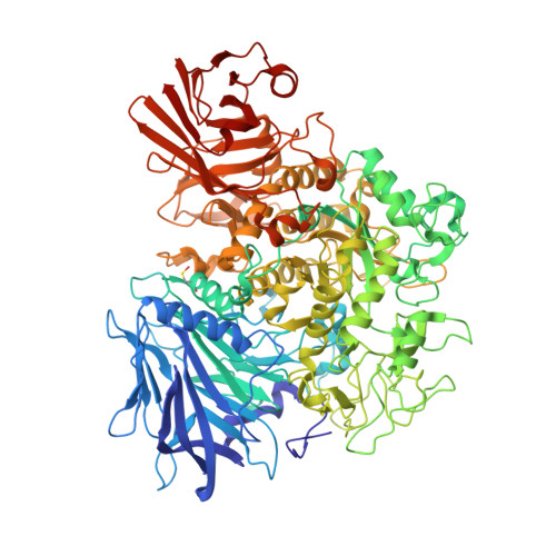
    α-1,4-Glucan lyase (EC 4.2.2.13) from the red seaweed Gracilariopsis lemaneiformis cleaves α-1,4-glucosidic linkages in glycogen, starch, and malto-oligosaccharides, yielding the keto-monosaccharide 1,5-anhydro-D-fructose. The enzyme belongs to glycoside hydrolase family 31 (GH31) but degrades starch via an elimination reaction instead of hydrolysis. The crystal structure shows that the enzyme, like GH31 hydrolases, contains a (β/α)8-barrel catalytic domain with B and B' subdomains, an N-terminal domain N, and the C-terminal domains C and D. The N-terminal domain N of the lyase was found to bind a trisaccharide. Complexes of the enzyme with acarbose and 1-dexoynojirimycin and two different covalent glycosyl-enzyme intermediates obtained with fluorinated sugar analogues show that, like GH31 hydrolases, the aspartic acid residues Asp(553) and Asp(665) are the catalytic nucleophile and acid, respectively. However, as a unique feature, the catalytic nucleophile is in a position to act also as a base that abstracts a proton from the C2 carbon atom of the covalently bound subsite -1 glucosyl residue, thus explaining the unique lyase activity of the enzyme. One Glu to Val mutation in the active site of the homologous α-glucosidase from Sulfolobus solfataricus resulted in a shift from hydrolytic to lyase activity, demonstrating that a subtle amino acid difference can promote lyase activity in a GH31 hydrolase.


  • Organizational Affiliation

    From the Laboratory of Biophysical Chemistry, Groningen Biomolecular Sciences and Biotechnology Institute, University of Groningen, Nijenborgh 7, 9747 AG Groningen, The Netherlands.


Macromolecules
Find similar proteins by:  (by identity cutoff)  |  3D Structure
Entity ID: 1
MoleculeChains Sequence LengthOrganismDetailsImage
ALPHA-1,4-GLUCAN LYASE ISOZYME 1
A, B, C, D
1,027Gracilariopsis lemaneiformisMutation(s): 0 
EC: 4.2.2.13
UniProt
Find proteins for Q9STC1 (Gracilariopsis lemaneiformis)
Explore Q9STC1 
Go to UniProtKB:  Q9STC1
Entity Groups  
Sequence Clusters30% Identity50% Identity70% Identity90% Identity95% Identity100% Identity
UniProt GroupQ9STC1
Sequence Annotations
Expand
  • Reference Sequence
Small Molecules
Ligands 3 Unique
IDChains Name / Formula / InChI Key2D Diagram3D Interactions
B9D
Query on B9D

Download Ideal Coordinates CCD File 
E [auth A],
J [auth B],
L [auth C]
5-fluoro-alpha-L-idopyranose
C6 H11 F O6
YQZCKDSOGGIGPL-DVKNGEFBSA-N
5DI
Query on 5DI

Download Ideal Coordinates CCD File 
O [auth D]alpha-D-threo-hexo-2,5-diulo-2,6-pyranosyl fluoride
C6 H9 F O5
VCJAHEOIRFFLTQ-SRQIZXRXSA-N
GOL
Query on GOL

Download Ideal Coordinates CCD File 
F [auth A]
G [auth A]
H [auth A]
I [auth A]
K [auth B]
GLYCEROL
C3 H8 O3
PEDCQBHIVMGVHV-UHFFFAOYSA-N
Modified Residues  1 Unique
IDChains TypeFormula2D DiagramParent
CSO
Query on CSO
A, B, C, D
L-PEPTIDE LINKINGC3 H7 N O3 SCYS
Experimental Data & Validation

Experimental Data

  • Method: X-RAY DIFFRACTION
  • Resolution: 1.90 Å
  • R-Value Free:  0.254 (Depositor), 0.260 (DCC) 
  • R-Value Work:  0.207 (Depositor), 0.210 (DCC) 
  • R-Value Observed: 0.209 (Depositor) 
Space Group: P 1
Unit Cell:
Length ( Å )Angle ( ˚ )
a = 92.1α = 80.31
b = 97.3β = 83.29
c = 136.28γ = 85.21
Software Package:
Software NamePurpose
REFMACrefinement
iMOSFLMdata reduction
SCALAdata scaling

Structure Validation

View Full Validation Report


Ligand Structure Quality Assessment 

Created with Raphaël 2.3.0Worse 01 BetterLigand structure goodness of fit to experimental dataBest fitted B9DClick on this verticalbar to view detailsBest fitted 5DIClick on this verticalbar to view details

Entry History 

Deposition Data

Revision History  (Full details and data files)

  • Version 1.0: 2013-03-27
    Type: Initial release
  • Version 1.1: 2013-08-14
    Changes: Database references, Refinement description
  • Version 1.2: 2013-09-25
    Changes: Database references
  • Version 1.3: 2019-01-30
    Changes: Data collection, Experimental preparation, Other
  • Version 1.4: 2019-02-06
    Changes: Data collection, Experimental preparation
  • Version 2.0: 2020-07-29
    Type: Remediation
    Reason: Carbohydrate remediation
    Changes: Atomic model, Derived calculations, Other, Structure summary
  • Version 2.1: 2023-12-20
    Changes: Data collection, Database references, Derived calculations, Refinement description, Structure summary
  • Version 2.2: 2024-10-09
    Changes: Structure summary