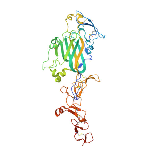
4AQT

Laminin gamma1 LN-LE1-2 structure


Experimental Data Snapshot

  • Method: X-RAY DIFFRACTION
  • Resolution: 3.20 Å
  • R-Value Free: 0.275 
  • R-Value Work: 0.231 
  • R-Value Observed: 0.231 

Starting Model: experimental
View more details

wwPDB Validation   3D Report Full Report


Ligand Structure Quality Assessment 


This is version 2.2 of the entry. See complete history


Literature

Crystal Structures of the Network-Forming Short-Arm Tips of the Laminin Beta1 and Gamma1 Chains.

Carafoli, F.Hussain, S.Hohenester, E.

(2012) PLoS One 7: 42473

  • DOI: https://doi.org/10.1371/journal.pone.0042473
  • Primary Citation of Related Structures:  
    4AQS, 4AQT

  • PubMed Abstract: 

    The heterotrimeric laminins are a defining component of basement membranes and essential for tissue formation and function in all animals. The three short arms of the cross-shaped laminin molecule are composed of one chain each and their tips mediate the formation of a polymeric network. The structural basis for laminin polymerisation is unknown. We have determined crystal structures of the short-arm tips of the mouse laminin β1 and γ1 chains, which are grossly similar to the previously determined structure of the corresponding α5 chain region. The short-arm tips consist of a laminin N-terminal (LN) domain that is attached like the head of a flower to a rod-like stem formed by tandem laminin-type epidermal growth factor-like (LE) domains. The LN domain is a β-sandwich with elaborate loop regions that differ between chains. The γ1 LN domain uniquely contains a calcium binding site. The LE domains have little regular structure and are stabilised by cysteines that are disulphide-linked 1-3, 2-4, 5-6 and 7-8 in all chains. The LN surface is not conserved across the α, β and γ chains, but within each chain subfamily there is a striking concentration of conserved residues on one face of the β-sandwich, while the opposite face invariably is shielded by glycans. We propose that the extensive conserved patches on the β and γ LN domains mediate the binding of these two chains to each other, and that the α chain LN domain subsequently binds to the composite β-γ surface. Mutations in the laminin β2 LN domain causing Pierson syndrome are likely to impair the folding of the β2 chain or its ability to form network interactions.


  • Organizational Affiliation

    Department of Life Sciences, Imperial College London, London, United Kingdom.


Macromolecules
Find similar proteins by:  (by identity cutoff)  |  3D Structure
Entity ID: 1
MoleculeChains Sequence LengthOrganismDetailsImage
LAMININ SUBUNIT GAMMA-1375Mus musculusMutation(s): 0 
UniProt
Find proteins for P02468 (Mus musculus)
Explore P02468 
Go to UniProtKB:  P02468
Entity Groups  
Sequence Clusters30% Identity50% Identity70% Identity90% Identity95% Identity100% Identity
UniProt GroupP02468
Glycosylation
Glycosylation Sites: 2Go to GlyGen: P02468-1
Sequence Annotations
Expand
  • Reference Sequence
Oligosaccharides

Help

Entity ID: 2
MoleculeChains Length2D Diagram Glycosylation3D Interactions
beta-D-mannopyranose-(1-4)-2-acetamido-2-deoxy-beta-D-glucopyranose-(1-4)-2-acetamido-2-deoxy-beta-D-glucopyranose
B
3N-Glycosylation
Glycosylation Resources
GlyTouCan:  G15407YE
GlyCosmos:  G15407YE
GlyGen:  G15407YE
Experimental Data & Validation

Experimental Data

  • Method: X-RAY DIFFRACTION
  • Resolution: 3.20 Å
  • R-Value Free: 0.275 
  • R-Value Work: 0.231 
  • R-Value Observed: 0.231 
  • Space Group: H 3 2
Unit Cell:
Length ( Å )Angle ( ˚ )
a = 203.01α = 90
b = 203.01β = 90
c = 93.69γ = 120
Software Package:
Software NamePurpose
CNSrefinement
XDSdata reduction
XDSdata scaling
PHASERphasing

Structure Validation

View Full Validation Report


Ligand Structure Quality Assessment 


Entry History 

Deposition Data

Revision History  (Full details and data files)

  • Version 1.0: 2012-08-15
    Type: Initial release
  • Version 2.0: 2020-07-29
    Type: Remediation
    Reason: Carbohydrate remediation
    Changes: Atomic model, Data collection, Derived calculations, Other, Structure summary
  • Version 2.1: 2023-12-20
    Changes: Data collection, Database references, Refinement description, Structure summary
  • Version 2.2: 2024-11-20
    Changes: Structure summary