4ARB

Mus musculus Acetylcholinesterase in complex with (S)-C5685 at 2.25 A resolution.


Experimental Data Snapshot

  • Method: X-RAY DIFFRACTION
  • Resolution: 2.25 Å
  • R-Value Free: 
    0.209 (Depositor), 0.200 (DCC) 
  • R-Value Work: 
    0.172 (Depositor), 0.160 (DCC) 
  • R-Value Observed: 
    0.172 (Depositor) 

Starting Model: experimental
View more details

wwPDB Validation   3D Report Full Report


Ligand Structure Quality Assessment 

Created with Raphaël 2.3.0Worse 01 BetterLigand structure goodness of fit to experimental dataBest fitted C57Click on this verticalbar to view detailsBest fitted NAGClick on this verticalbar to view details

This is version 2.4 of the entry. See complete history


Literature

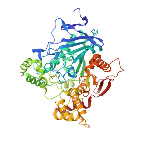
Similar But Different: Thermodynamic and Structural Characterization of a Pair of Enantiomers Binding to Acetylcholinesterase.

Berg, L.Niemiec, M.S.Qian, W.Andersson, C.D.Wittung-Stafshede, P.Ekstrom, F.Linusson, A.

(2012) Angew Chem Int Ed Engl 51: 12716


Macromolecules
Find similar proteins by:  (by identity cutoff)  |  3D Structure
Entity ID: 1
MoleculeChains Sequence LengthOrganismDetailsImage
ACETYLCHOLINESTERASE
A, B
548Mus musculusMutation(s): 0 
EC: 3.1.1.7
UniProt & NIH Common Fund Data Resources
Find proteins for P21836 (Mus musculus)
Explore P21836 
Go to UniProtKB:  P21836
IMPC:  MGI:87876
Entity Groups  
Sequence Clusters30% Identity50% Identity70% Identity90% Identity95% Identity100% Identity
UniProt GroupP21836
Glycosylation
Glycosylation Sites: 2Go to GlyGen: P21836-1
Sequence Annotations
Expand
  • Reference Sequence
Small Molecules
Ligands 4 Unique
IDChains Name / Formula / InChI Key2D Diagram3D Interactions
C57
Query on C57

Download Ideal Coordinates CCD File 
C [auth A],
J [auth B]
4-(DIMETHYLAMINO)-N-{[(2S)-1-ETHYLPYRROLIDIN-2-YL]METHYL}-2-METHOXY-5-NITROBENZAMIDE
C17 H26 N4 O4
AFNPLUMDSLEAPD-LBPRGKRZSA-N
P6G
Query on P6G

Download Ideal Coordinates CCD File 
K [auth B]HEXAETHYLENE GLYCOL
C12 H26 O7
IIRDTKBZINWQAW-UHFFFAOYSA-N
NAG
Query on NAG

Download Ideal Coordinates CCD File 
E [auth A],
G [auth A],
M [auth B]
2-acetamido-2-deoxy-beta-D-glucopyranose
C8 H15 N O6
OVRNDRQMDRJTHS-FMDGEEDCSA-N
PEG
Query on PEG

Download Ideal Coordinates CCD File 
D [auth A]
F [auth A]
H [auth A]
I [auth A]
L [auth B]
DI(HYDROXYETHYL)ETHER
C4 H10 O3
MTHSVFCYNBDYFN-UHFFFAOYSA-N
Experimental Data & Validation

Experimental Data

  • Method: X-RAY DIFFRACTION
  • Resolution: 2.25 Å
  • R-Value Free:  0.209 (Depositor), 0.200 (DCC) 
  • R-Value Work:  0.172 (Depositor), 0.160 (DCC) 
  • R-Value Observed: 0.172 (Depositor) 
Space Group: P 21 21 21
Unit Cell:
Length ( Å )Angle ( ˚ )
a = 78.926α = 90
b = 111.781β = 90
c = 227.071γ = 90
Software Package:
Software NamePurpose
PHENIXrefinement
XDSdata reduction
SCALAdata scaling
REFMACphasing

Structure Validation

View Full Validation Report


Ligand Structure Quality Assessment 

Created with Raphaël 2.3.0Worse 01 BetterLigand structure goodness of fit to experimental dataBest fitted C57Click on this verticalbar to view detailsBest fitted NAGClick on this verticalbar to view details

Entry History 

Deposition Data

Revision History  (Full details and data files)

  • Version 1.0: 2012-11-28
    Type: Initial release
  • Version 1.1: 2013-01-16
    Changes: Database references
  • Version 2.0: 2018-01-17
    Changes: Atomic model, Data collection
  • Version 2.1: 2018-02-28
    Changes: Source and taxonomy
  • Version 2.2: 2020-07-29
    Type: Remediation
    Reason: Carbohydrate remediation
    Changes: Data collection, Derived calculations, Other, Structure summary
  • Version 2.3: 2023-12-20
    Changes: Data collection, Database references, Refinement description, Structure summary
  • Version 2.4: 2024-11-06
    Changes: Structure summary