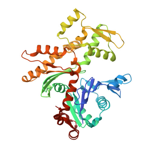
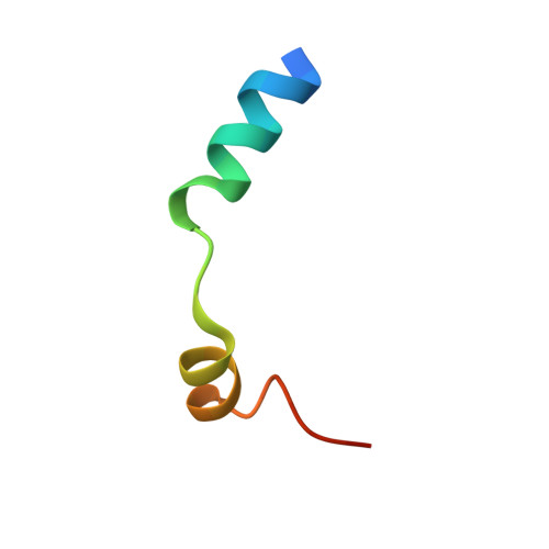
4B1X

Structure of the Phactr1 RPEL-2 bound to G-actin


Experimental Data Snapshot

  • Method: X-RAY DIFFRACTION
  • Resolution: 1.80 Å
  • R-Value Free: 0.213 
  • R-Value Work: 0.169 
  • R-Value Observed: 0.171 

Starting Model: experimental
View more details

wwPDB Validation   3D Report Full Report


Ligand Structure Quality Assessment 


This is version 1.5 of the entry. See complete history


Literature

Structures of the Phactr1 RPEL domain and RPEL motif complexes with G-actin reveal the molecular basis for actin binding cooperativity.

Mouilleron, S.Wiezlak, M.O'Reilly, N.Treisman, R.McDonald, N.Q.

(2012) Structure 20: 1960-1970

  • DOI: https://doi.org/10.1016/j.str.2012.08.031
  • Primary Citation of Related Structures:  
    4B1U, 4B1V, 4B1W, 4B1X, 4B1Y, 4B1Z

  • PubMed Abstract: 

    The Phactr family of PP1-binding proteins and the myocardin-related transcription factor family of transcriptional coactivators contain regulatory domains comprising three copies of the RPEL motif, a G-actin binding element. We report the structure of a Phactr1 G-actin⋅RPEL domain complex. Three G-actins surround the crank-shaped RPEL domain forming a closed helical assembly. Their spatial relationship is identical to the RPEL-actins within the pentavalent MRTF G-actin⋅RPEL domain complex, suggesting that conserved cooperative interactions between actin⋅RPEL units organize the assembly. In the trivalent Phactr1 complex, each G-actin⋅RPEL unit makes secondary contacts with its downstream actin involving distinct RPEL residues. Similar secondary contacts are seen in G-actin⋅RPEL peptide crystals. Loss-of-secondary-contact mutations destabilize the Phactr1 G-actin⋅RPEL assembly. Furthermore, actin-mediated inhibition of Phactr1 nuclear import requires secondary contact residues in the Phactr1 N-terminal RPEL-N motif, suggesting that it involves interaction of RPEL-N with the C-terminal assembly. Secondary actin contacts by actin-bound RPEL motifs thus govern formation of multivalent actin⋅RPEL assemblies.


  • Organizational Affiliation

    Structural Biology, CRUK London Research Institute, 44 Lincoln's Inn Fields, London WC2A 3LY, UK.


Macromolecules
Find similar proteins by:  (by identity cutoff)  |  3D Structure
Entity ID: 1
MoleculeChains Sequence LengthOrganismDetailsImage
ACTIN, ALPHA SKELETAL MUSCLEA [auth B]376Oryctolagus cuniculusMutation(s): 0 
EC: 3.6.4
UniProt
Find proteins for P68135 (Oryctolagus cuniculus)
Explore P68135 
Go to UniProtKB:  P68135
Entity Groups  
Sequence Clusters30% Identity50% Identity70% Identity90% Identity95% Identity100% Identity
UniProt GroupP68135
Sequence Annotations
Expand
  • Reference Sequence
Find similar proteins by:  (by identity cutoff)  |  3D Structure
Entity ID: 2
MoleculeChains Sequence LengthOrganismDetailsImage
PHOSPHATASE AND ACTIN REGULATOR 1B [auth M]32Mus musculusMutation(s): 0 
UniProt & NIH Common Fund Data Resources
Find proteins for Q2M3X8 (Mus musculus)
Explore Q2M3X8 
Go to UniProtKB:  Q2M3X8
IMPC:  MGI:2659021
Entity Groups  
Sequence Clusters30% Identity50% Identity70% Identity90% Identity95% Identity100% Identity
UniProt GroupQ2M3X8
Sequence Annotations
Expand
  • Reference Sequence
Experimental Data & Validation

Experimental Data

  • Method: X-RAY DIFFRACTION
  • Resolution: 1.80 Å
  • R-Value Free: 0.213 
  • R-Value Work: 0.169 
  • R-Value Observed: 0.171 
  • Space Group: P 32 2 1
Unit Cell:
Length ( Å )Angle ( ˚ )
a = 77.28α = 90
b = 77.28β = 90
c = 130.809γ = 120
Software Package:
Software NamePurpose
PHENIXrefinement
PHASERphasing

Structure Validation

View Full Validation Report


Ligand Structure Quality Assessment 


Entry History 

Deposition Data

Revision History  (Full details and data files)

  • Version 1.0: 2013-07-31
    Type: Initial release
  • Version 1.1: 2014-10-22
    Changes: Database references
  • Version 1.2: 2018-02-28
    Changes: Database references
  • Version 1.3: 2018-04-25
    Changes: Data collection
  • Version 1.4: 2018-06-20
    Changes: Data collection, Derived calculations
  • Version 1.5: 2023-12-20
    Changes: Data collection, Database references, Derived calculations, Other, Refinement description