4BCM

Structure of CDK2 in complex with cyclin A and a 2-amino-4-heteroaryl- pyrimidine inhibitor


Experimental Data Snapshot

  • Method: X-RAY DIFFRACTION
  • Resolution: 2.45 Å
  • R-Value Free: 
    0.258 (Depositor) 
  • R-Value Work: 
    0.196 (Depositor), 0.188 (DCC) 
  • R-Value Observed: 
    0.198 (Depositor) 

wwPDB Validation   3D Report Full Report


Ligand Structure Quality Assessment 

Created with Raphaël 2.3.0Worse 01 BetterLigand structure goodness of fit to experimental dataBest fitted T7ZClick on this verticalbar to view details

This is version 1.1 of the entry. See complete history


Literature

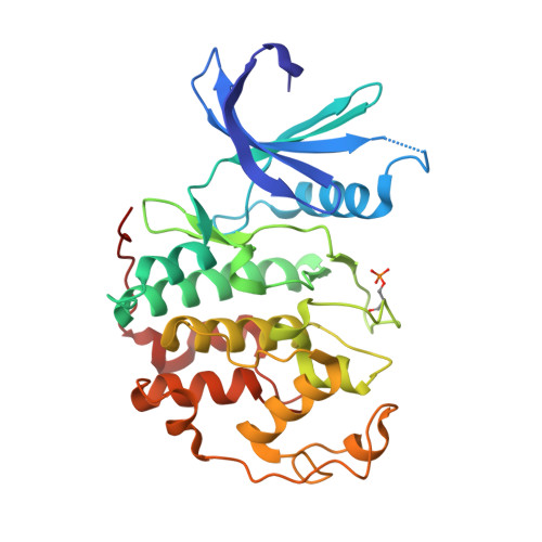
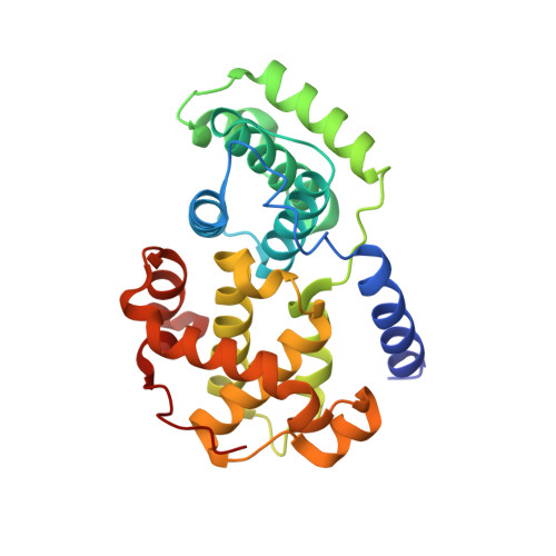
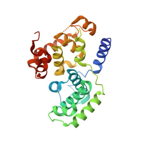
Comparative Structural and Functional Studies of 4-(Thiazol- 5-Yl)-2-(Phenylamino)Pyrimidine-5-Carbonitrile Cdk9 Inhibitors Suggest the Basis for Isotype Selectivity.

Hole, A.J.Baumli, S.Shao, H.Shi, S.Pepper, C.Fischer, P.M.Wang, S.Endicott, J.A.Noble, M.E.M.

(2013) J Med Chem 56: 660

  • DOI: https://doi.org/10.1021/jm301495v
  • Primary Citation of Related Structures:  
    4BCF, 4BCH, 4BCI, 4BCJ, 4BCK, 4BCM, 4BCN, 4BCO, 4BCQ

  • PubMed Abstract: 

    Cyclin-dependent kinase 9/cyclin T, the protein kinase heterodimer that constitutes positive transcription elongation factor b, is a well-validated target for treatment of several diseases, including cancer and cardiac hypertrophy. In order to aid inhibitor design and rationalize the basis for CDK9 selectivity, we have studied the CDK-binding properties of six different members of a 4-(thiazol-5-yl)-2-(phenylamino)pyrimidine-5-carbonitrile series that bind to both CDK9/cyclin T and CDK2/cyclin A. We find that for a given CDK, the melting temperature of a CDK/cyclin/inhibitor complex correlates well with inhibitor potency, suggesting that differential scanning fluorimetry (DSF) is a useful orthogonal measure of inhibitory activity for this series. We have used DSF to demonstrate that the binding of these compounds is independent of the presence or absence of the C-terminal tail region of CDK9, unlike the binding of the CDK9-selective inhibitor 5,6-dichlorobenzimidazone-1-β-d-ribofuranoside (DRB). Finally, on the basis of 11 cocrystal structures bound to CDK9/cyclin T or CDK2/cyclin A, we conclude that selective inhibition of CDK9/cyclin T by members of the 4-(thiazol-5-yl)-2-(phenylamino)pyrimidine-5-carbonitrile series results from the relative malleability of the CDK9 active site rather than from the formation of specific polar contacts.


  • Organizational Affiliation

    Department of Biochemistry, University of Oxford, South Parks Road, Oxford OX1 3QU, UK.


Macromolecules
Find similar proteins by:  (by identity cutoff)  |  3D Structure
Entity ID: 1
MoleculeChains Sequence LengthOrganismDetailsImage
CYCLIN-DEPENDENT KINASE 2
A, C
301Homo sapiensMutation(s): 0 
EC: 2.7.11.22
UniProt & NIH Common Fund Data Resources
Find proteins for P24941 (Homo sapiens)
Explore P24941 
Go to UniProtKB:  P24941
PHAROS:  P24941
GTEx:  ENSG00000123374 
Entity Groups  
Sequence Clusters30% Identity50% Identity70% Identity90% Identity95% Identity100% Identity
UniProt GroupP24941
Sequence Annotations
Expand
  • Reference Sequence
Find similar proteins by:  (by identity cutoff)  |  3D Structure
Entity ID: 2
MoleculeChains Sequence LengthOrganismDetailsImage
CYCLIN-A2262Homo sapiensMutation(s): 0 
UniProt & NIH Common Fund Data Resources
Find proteins for P20248 (Homo sapiens)
Explore P20248 
Go to UniProtKB:  P20248
PHAROS:  P20248
GTEx:  ENSG00000145386 
Entity Groups  
Sequence Clusters30% Identity50% Identity70% Identity90% Identity95% Identity100% Identity
UniProt GroupP20248
Sequence Annotations
Expand
  • Reference Sequence
Find similar proteins by:  (by identity cutoff)  |  3D Structure
Entity ID: 3
MoleculeChains Sequence LengthOrganismDetailsImage
CYCLIN-A2262Homo sapiensMutation(s): 0 
UniProt & NIH Common Fund Data Resources
Find proteins for P20248 (Homo sapiens)
Explore P20248 
Go to UniProtKB:  P20248
PHAROS:  P20248
GTEx:  ENSG00000145386 
Entity Groups  
Sequence Clusters30% Identity50% Identity70% Identity90% Identity95% Identity100% Identity
UniProt GroupP20248
Sequence Annotations
Expand
  • Reference Sequence
Small Molecules
Modified Residues  1 Unique
IDChains TypeFormula2D DiagramParent
TPO
Query on TPO
A, C
L-PEPTIDE LINKINGC4 H10 N O6 PTHR
Experimental Data & Validation

Experimental Data

  • Method: X-RAY DIFFRACTION
  • Resolution: 2.45 Å
  • R-Value Free:  0.258 (Depositor) 
  • R-Value Work:  0.196 (Depositor), 0.188 (DCC) 
  • R-Value Observed: 0.198 (Depositor) 
Space Group: P 21 21 21
Unit Cell:
Length ( Å )Angle ( ˚ )
a = 74.07α = 90
b = 135.41β = 90
c = 148.63γ = 90
Software Package:
Software NamePurpose
PHENIXrefinement
PHASERphasing

Structure Validation

View Full Validation Report


Ligand Structure Quality Assessment 

Created with Raphaël 2.3.0Worse 01 BetterLigand structure goodness of fit to experimental dataBest fitted T7ZClick on this verticalbar to view details

Entry History 

Deposition Data

Revision History  (Full details and data files)

  • Version 1.0: 2013-03-06
    Type: Initial release
  • Version 1.1: 2024-10-23
    Changes: Data collection, Database references, Derived calculations, Other, Structure summary