4BW5 | pdb_00004bw5

Crystal structure of human two pore domain potassium ion channel TREK2 (K2P10.1)


Experimental Data Snapshot

  • Method: X-RAY DIFFRACTION
  • Resolution: 3.20 Å
  • R-Value Free: 
    0.254 (Depositor), 0.272 (DCC) 
  • R-Value Work: 
    0.237 (Depositor), 0.252 (DCC) 
  • R-Value Observed: 
    0.238 (Depositor) 

Starting Models: experimental
View more details

wwPDB Validation 3D Report Full Report

Validation slider image for 4BW5

Ligand Structure Quality Assessment 


This is version 1.5 of the entry. See complete history

Literature

K2P Channel Gating Mechanisms Revealed by Structures of Trek-2 and a Complex with Prozac

Pike, A.C.W.Mackenzie, A.Mcclenaghan, C.Aryal, P.Dong, L.Quigley, A.Grieben, M.Goubin, S.Mukhopadhyay, S.Ruda, G.F.Clausen, M.V.Cao, L.Brennan, P.E.Burgess-Brown, N.A.Sansom, M.S.P.Tucker, S.J.Carpenter, E.P.

(2015) Science 347: 1256

  • DOI: https://doi.org/10.1126/science.1261512
  • Primary Citation Related Structures: 
    4BW5, 4XDJ, 4XDK, 4XDL

  • PubMed Abstract: 

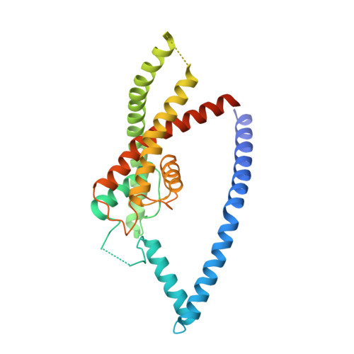
    TREK-2 (KCNK10/K2P10), a two-pore domain potassium (K2P) channel, is gated by multiple stimuli such as stretch, fatty acids, and pH and by several drugs. However, the mechanisms that control channel gating are unclear. Here we present crystal structures of the human TREK-2 channel (up to 3.4 angstrom resolution) in two conformations and in complex with norfluoxetine, the active metabolite of fluoxetine (Prozac) and a state-dependent blocker of TREK channels. Norfluoxetine binds within intramembrane fenestrations found in only one of these two conformations. Channel activation by arachidonic acid and mechanical stretch involves conversion between these states through movement of the pore-lining helices. These results provide an explanation for TREK channel mechanosensitivity, regulation by diverse stimuli, and possible off-target effects of the serotonin reuptake inhibitor Prozac.


  • Organizational Affiliation
    • Structural Genomics Consortium, University of Oxford, Oxford OX3 7DQ, UK.

Macromolecule Content 

  • Total Structure Weight: 126.36 kDa 
  • Atom Count: 7,235 
  • Modeled Residue Count: 985 
  • Deposited Residue Count: 1,128 
  • Unique protein chains: 1

Macromolecules

Find similar proteins by:|  3D Structure
Entity ID: 1
MoleculeChains  Sequence LengthOrganismDetailsImage
POTASSIUM CHANNEL SUBFAMILY K MEMBER 10
A, B, C, D
282Homo sapiensMutation(s): 0 
Membrane Entity: Yes 
UniProt & NIH Common Fund Data Resources
Find proteins for P57789 (Homo sapiens)
Explore P57789 
Go to UniProtKB:  P57789
PHAROS:  P57789
GTEx:  ENSG00000100433 
Entity Groups
Sequence Clusters30% Identity50% Identity70% Identity90% Identity95% Identity100% Identity
UniProt GroupP57789
Sequence Annotations
Expand
Reference Sequence

Small Molecules

Ligands 3 Unique
IDChains Name / Formula / InChI Key2D Diagram3D Interactions
PC1

Query on PC1


Download:Ideal Coordinates CCD File
K [auth A],
L [auth B]
1,2-DIACYL-SN-GLYCERO-3-PHOSPHOCHOLINE
C44 H88 N O8 P
NRJAVPSFFCBXDT-HUESYALOSA-N
CD

Query on CD


Download:Ideal Coordinates CCD File
I [auth A],
J [auth A]
CADMIUM ION
Cd
WLZRMCYVCSSEQC-UHFFFAOYSA-N
K

Query on K


Download:Ideal Coordinates CCD File
E [auth A]
F [auth A]
G [auth A]
H [auth A]
M [auth C]
E [auth A],
F [auth A],
G [auth A],
H [auth A],
M [auth C],
N [auth C],
O [auth C],
P [auth C]
POTASSIUM ION
K
NPYPAHLBTDXSSS-UHFFFAOYSA-N

Experimental Data & Validation

Experimental Data

  • Method: X-RAY DIFFRACTION
  • Resolution: 3.20 Å
  • R-Value Free:  0.254 (Depositor), 0.272 (DCC) 
  • R-Value Work:  0.237 (Depositor), 0.252 (DCC) 
  • R-Value Observed: 0.238 (Depositor) 
Space Group: P 1 21 1
Unit Cell:
Length ( Å )Angle ( ˚ )
a = 87.83α = 90
b = 96.96β = 92.58
c = 103.7γ = 90
Software Package:
Software NamePurpose
BUSTERrefinement
XDSdata reduction
Aimlessdata scaling
PHASERphasing

Structure Validation

View Full Validation Report


Ligand Structure Quality Assessment 


Entry History 

Revision History  (Full details and data files)

  • Version 1.0: 2013-07-31
    Type: Initial release
  • Version 1.1: 2015-03-18
    Changes: Database references
  • Version 1.2: 2015-03-25
    Changes: Database references
  • Version 1.3: 2019-03-06
    Changes: Data collection, Experimental preparation, Other
  • Version 1.4: 2023-12-20
    Changes: Data collection, Database references, Derived calculations, Other, Refinement description
  • Version 1.5: 2024-11-06
    Changes: Structure summary