4C0S | pdb_00004c0s

Mammalian translation elongation factor eEF1A2


Experimental Data Snapshot

  • Method: X-RAY DIFFRACTION
  • Resolution: 2.70 Å
  • R-Value Free: 
    0.256 (Depositor), 0.255 (DCC) 
  • R-Value Work: 
    0.202 (Depositor), 0.203 (DCC) 
  • R-Value Observed: 
    0.205 (Depositor) 

wwPDB Validation 3D Report Full Report

Validation slider image for 4C0S

Ligand Structure Quality Assessment 


This is version 1.4 of the entry. See complete history

Literature

Mammalian Translation Elongation Factor Eef1A2: X-Ray Structure and New Features of Gdp/GTP Exchange Mechanism in Higher Eukaryotes

Crepin, T.Mccarthy, A.Negrutskii, A.Shalak, V.F.Tukalo, V.Vlasenko, D.O.Yaremchuk, A.D.

(2014) Nucleic Acids Res 42: 12939

  • DOI: https://doi.org/10.1093/nar/gku974
  • Primary Citation Related Structures: 
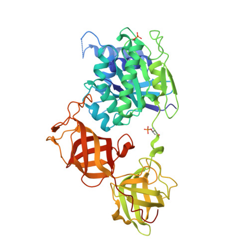
    4C0S

  • PubMed Abstract: 

    Eukaryotic elongation factor eEF1A transits between the GTP- and GDP-bound conformations during the ribosomal polypeptide chain elongation. eEF1A*GTP establishes a complex with the aminoacyl-tRNA in the A site of the 80S ribosome. Correct codon-anticodon recognition triggers GTP hydrolysis, with subsequent dissociation of eEF1A*GDP from the ribosome. The structures of both the 'GTP'- and 'GDP'-bound conformations of eEF1A are unknown. Thus, the eEF1A-related ribosomal mechanisms were anticipated only by analogy with the bacterial homolog EF-Tu. Here, we report the first crystal structure of the mammalian eEF1A2*GDP complex which indicates major differences in the organization of the nucleotide-binding domain and intramolecular movements of eEF1A compared to EF-Tu. Our results explain the nucleotide exchange mechanism in the mammalian eEF1A and suggest that the first step of eEF1A*GDP dissociation from the 80S ribosome is the rotation of the nucleotide-binding domain observed after GTP hydrolysis.


  • Organizational Affiliation
    • University of Grenoble Alpes, UVHCI, F-38000 Grenoble, France CNRS, UVHCI, F-38000 Grenoble, France Unit for Virus Host-Cell Interactions, University of Grenoble Alpes-EMBL-CNRS, 71 avenue des Martyrs, 38042 France.

Macromolecule Content 

  • Total Structure Weight: 102.35 kDa 
  • Atom Count: 6,951 
  • Modeled Residue Count: 893 
  • Deposited Residue Count: 926 
  • Unique protein chains: 1

Macromolecules

Find similar proteins by:|  3D Structure
Entity ID: 1
MoleculeChains  Sequence LengthOrganismDetailsImage
ELONGATION FACTOR 1-ALPHA 2
A, B
463Oryctolagus cuniculusMutation(s): 0 
EC: 3.6.5
UniProt
Find proteins for Q71V39 (Oryctolagus cuniculus)
Explore Q71V39 
Go to UniProtKB:  Q71V39
Entity Groups
Sequence Clusters30% Identity50% Identity70% Identity90% Identity95% Identity100% Identity
UniProt GroupQ71V39
Sequence Annotations
Expand
Reference Sequence

Experimental Data & Validation

Experimental Data

  • Method: X-RAY DIFFRACTION
  • Resolution: 2.70 Å
  • R-Value Free:  0.256 (Depositor), 0.255 (DCC) 
  • R-Value Work:  0.202 (Depositor), 0.203 (DCC) 
  • R-Value Observed: 0.205 (Depositor) 
Space Group: P 61 2 2
Unit Cell:
Length ( Å )Angle ( ˚ )
a = 135.37α = 90
b = 135.37β = 90
c = 304.62γ = 120
Software Package:
Software NamePurpose
REFMACrefinement
XDSdata reduction
XSCALEdata scaling

Structure Validation

View Full Validation Report


Ligand Structure Quality Assessment 


Entry History 

Deposition Data

Revision History  (Full details and data files)

  • Version 1.0: 2014-08-27
    Type: Initial release
  • Version 1.1: 2014-10-29
    Changes: Database references
  • Version 1.2: 2014-11-26
    Changes: Database references
  • Version 1.3: 2017-01-25
    Changes: Data collection
  • Version 1.4: 2024-11-20
    Changes: Data collection, Database references, Derived calculations, Other, Structure summary