4D90 | pdb_00004d90

Crystal Structure of Del-1 EGF domains


Experimental Data Snapshot

  • Method: X-RAY DIFFRACTION
  • Resolution: 2.60 Å
  • R-Value Free: 
    0.259 (Depositor), 0.257 (DCC) 
  • R-Value Work: 
    0.204 (Depositor), 0.206 (DCC) 
  • R-Value Observed: 
    0.207 (Depositor) 

Starting Model: experimental
View more details

wwPDB Validation 3D Report Full Report

Validation slider image for 4D90

Ligand Structure Quality Assessment 


This is version 1.5 of the entry. See complete history

Literature

The RGD finger of Del-1 is a unique structural feature critical for integrin binding.

Schurpf, T.Chen, Q.Liu, J.H.Wang, R.Springer, T.A.Wang, J.H.

(2012) FASEB J 26: 3412-3420

  • DOI: https://doi.org/10.1096/fj.11-202036
  • Primary Citation Related Structures: 
    4D90

  • PubMed Abstract: 

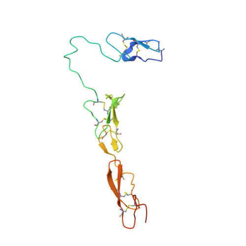
    Developmental endothelial cell locus-1 (Del-1) glycoprotein is secreted by endothelial cells and a subset of macrophages. Del-1 plays a regulatory role in vascular remodeling and functions in innate immunity through interaction with integrin α(V)β(3). Del-1 contains 3 epidermal growth factor (EGF)-like repeats and 2 discoidin-like domains. An Arg-Gly-Asp (RGD) motif in the second EGF domain (EGF2) mediates adhesion by endothelial cells and phagocytes. We report the crystal structure of its 3 EGF domains. The RGD motif of EGF2 forms a type II' β turn at the tip of a long protruding loop, dubbed the RGD finger. Whereas EGF2 and EGF3 constitute a rigid rod via an interdomain calcium ion binding site, the long linker between EGF1 and EGF2 lends considerable flexibility to EGF1. Two unique O-linked glycans and 1 N-linked glycan locate to the opposite side of EGF2 from the RGD motif. These structural features favor integrin binding of the RGD finger. Mutagenesis data confirm the importance of having the RGD motif at the tip of the RGD finger. A database search for EGF domain sequences shows that this RGD finger is likely an evolutionary insertion and unique to the EGF domain of Del-1 and its homologue milk fat globule-EGF 8.


  • Organizational Affiliation
    • Immune Disease Institute, Harvard Medical School, Boston, MA, USA.

Macromolecule Content 

  • Total Structure Weight: 31.78 kDa 
  • Atom Count: 1,994 
  • Modeled Residue Count: 253 
  • Deposited Residue Count: 286 
  • Unique protein chains: 1

Macromolecules

Find similar proteins by:|  3D Structure
Entity ID: 1
MoleculeChains  Sequence LengthOrganismDetailsImage
EGF-like repeat and discoidin I-like domain-containing protein 3
A, B
143Homo sapiensMutation(s): 0 
Gene Names: del-1DEL1EDIL3
UniProt & NIH Common Fund Data Resources
Find proteins for O43854 (Homo sapiens)
Explore O43854 
Go to UniProtKB:  O43854
PHAROS:  O43854
GTEx:  ENSG00000164176 
Entity Groups
Sequence Clusters30% Identity50% Identity70% Identity90% Identity95% Identity100% Identity
UniProt GroupO43854
Glycosylation
Glycosylation Sites: 3Go to GlyGen: O43854-1
Sequence Annotations
Expand
Reference Sequence

Small Molecules

Ligands 4 Unique
IDChains Name / Formula / InChI Key2D Diagram3D Interactions
NAG

Query on NAG


Download:Ideal Coordinates CCD File
D [auth A],
H [auth B]
2-acetamido-2-deoxy-beta-D-glucopyranose
C8 H15 N O6
OVRNDRQMDRJTHS-FMDGEEDCSA-N
NGA

Query on NGA


Download:Ideal Coordinates CCD File
C [auth A],
G [auth B]
2-acetamido-2-deoxy-beta-D-galactopyranose
C8 H15 N O6
OVRNDRQMDRJTHS-JAJWTYFOSA-N
FUC

Query on FUC


Download:Ideal Coordinates CCD File
E [auth A],
I [auth B]
alpha-L-fucopyranose
C6 H12 O5
SHZGCJCMOBCMKK-SXUWKVJYSA-N
CA

Query on CA


Download:Ideal Coordinates CCD File
F [auth A],
J [auth B]
CALCIUM ION
Ca
BHPQYMZQTOCNFJ-UHFFFAOYSA-N

Experimental Data & Validation

Experimental Data

  • Method: X-RAY DIFFRACTION
  • Resolution: 2.60 Å
  • R-Value Free:  0.259 (Depositor), 0.257 (DCC) 
  • R-Value Work:  0.204 (Depositor), 0.206 (DCC) 
  • R-Value Observed: 0.207 (Depositor) 
Space Group: P 32
Unit Cell:
Length ( Å )Angle ( ˚ )
a = 82.68α = 90
b = 82.68β = 90
c = 56.422γ = 120
Software Package:
Software NamePurpose
DENZOdata reduction
SCALEPACKdata scaling
PHASERphasing
PHENIXrefinement
PDB_EXTRACTdata extraction
HKL-2000data collection
HKL-2000data reduction
HKL-2000data scaling

Structure Validation

View Full Validation Report


Ligand Structure Quality Assessment 


Entry History 

Deposition Data

Revision History  (Full details and data files)

  • Version 1.0: 2012-05-30
    Type: Initial release
  • Version 1.1: 2013-01-02
    Changes: Database references
  • Version 1.2: 2017-11-15
    Changes: Refinement description
  • Version 1.3: 2020-07-29
    Type: Remediation
    Reason: Carbohydrate remediation
    Changes: Data collection, Database references, Derived calculations, Structure summary
  • Version 1.4: 2023-09-13
    Changes: Data collection, Database references, Refinement description, Structure summary
  • Version 1.5: 2024-11-20
    Changes: Structure summary