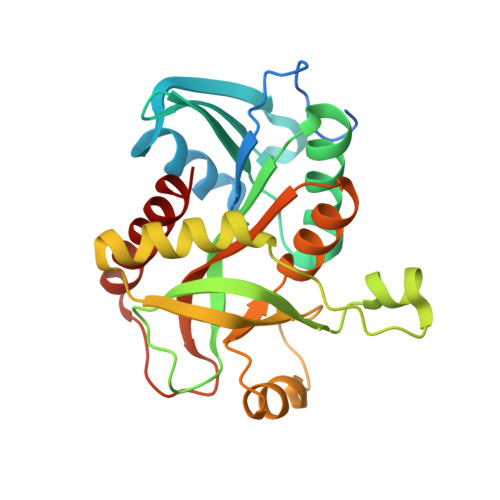
Insights into phosphate cooperativity and influence of substrate modifications on binding and catalysis of hexameric purine nucleoside phosphorylases.
de Giuseppe, P.O., Martins, N.H., Meza, A.N., Dos Santos, C.R., Pereira, H.D., Murakami, M.T.(2012) PLoS One 7: e44282-e44282
- PubMed: 22957058
- DOI: https://doi.org/10.1371/journal.pone.0044282
- Primary Citation of Related Structures:
4D8V, 4D8X, 4D8Y, 4D98, 4D9H, 4DA0, 4DA6, 4DA7, 4DA8, 4DAB, 4DAE, 4DAN, 4DAO, 4DAR - PubMed Abstract:
The hexameric purine nucleoside phosphorylase from Bacillus subtilis (BsPNP233) displays great potential to produce nucleoside analogues in industry and can be exploited in the development of new anti-tumor gene therapies. In order to provide structural basis for enzyme and substrates rational optimization, aiming at those applications, the present work shows a thorough and detailed structural description of the binding mode of substrates and nucleoside analogues to the active site of the hexameric BsPNP233. Here we report the crystal structure of BsPNP233 in the apo form and in complex with 11 ligands, including clinically relevant compounds. The crystal structure of six ligands (adenine, 2'deoxyguanosine, aciclovir, ganciclovir, 8-bromoguanosine, 6-chloroguanosine) in complex with a hexameric PNP are presented for the first time. Our data showed that free bases adopt alternative conformations in the BsPNP233 active site and indicated that binding of the co-substrate (2'deoxy)ribose 1-phosphate might contribute for stabilizing the bases in a favorable orientation for catalysis. The BsPNP233-adenosine complex revealed that a hydrogen bond between the 5' hydroxyl group of adenosine and Arg(43*) side chain contributes for the ribosyl radical to adopt an unusual C3'-endo conformation. The structures with 6-chloroguanosine and 8-bromoguanosine pointed out that the Cl(6) and Br(8) substrate modifications seem to be detrimental for catalysis and can be explored in the design of inhibitors for hexameric PNPs from pathogens. Our data also corroborated the competitive inhibition mechanism of hexameric PNPs by tubercidin and suggested that the acyclic nucleoside ganciclovir is a better inhibitor for hexameric PNPs than aciclovir. Furthermore, comparative structural analyses indicated that the replacement of Ser(90) by a threonine in the B. cereus hexameric adenosine phosphorylase (Thr(91)) is responsible for the lack of negative cooperativity of phosphate binding in this enzyme.
- Laboratório Nacional de Biociências, Centro Nacional de Pesquisa em Energia e Materiais, Campinas, São Paulo, Brazil.
Organizational Affiliation:


