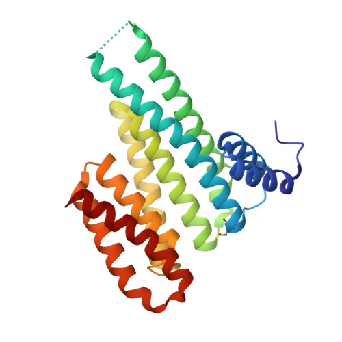
4DHU

Small-molecule inhibitors of 14-3-3 protein-protein interactions from virtual screening


Experimental Data Snapshot

  • Method: X-RAY DIFFRACTION
  • Resolution: 1.67 Å
  • R-Value Free: 0.176 
  • R-Value Work: 0.131 
  • R-Value Observed: 0.133 

wwPDB Validation   3D Report Full Report


Ligand Structure Quality Assessment 


This is version 1.2 of the entry. See complete history


Literature

Virtual screening and experimental validation reveal novel small-molecule inhibitors of 14-3-3 protein-protein interactions.

Thiel, P.Roglin, L.Meissner, N.Hennig, S.Kohlbacher, O.Ottmann, C.

(2013) Chem Commun (Camb) 49: 8468-8470

  • DOI: https://doi.org/10.1039/c3cc44612c
  • Primary Citation of Related Structures:  
    3T0L, 3T0M, 4DHM, 4DHN, 4DHO, 4DHP, 4DHQ, 4DHR, 4DHS, 4DHT, 4DHU

  • PubMed Abstract: 

    We report first non-covalent and exclusively extracellular inhibitors of 14-3-3 protein-protein interactions identified by virtual screening. Optimization by crystal structure analysis and in vitro binding assays yielded compounds capable of disrupting the interaction of 14-3-3σ with aminopeptidase N in a cellular assay.


  • Organizational Affiliation

    Chemical Genomics Centre of the Max Planck Society, Otto-Hahn-Straße 15, D-44227 Dortmund, Germany.


Macromolecules
Find similar proteins by:  (by identity cutoff)  |  3D Structure
Entity ID: 1
MoleculeChains Sequence LengthOrganismDetailsImage
14-3-3 PROTEIN SIGMA235Homo sapiensMutation(s): 0 
Gene Names: HME1SFN
UniProt & NIH Common Fund Data Resources
Find proteins for P31947 (Homo sapiens)
Explore P31947 
Go to UniProtKB:  P31947
PHAROS:  P31947
GTEx:  ENSG00000175793 
Entity Groups  
Sequence Clusters30% Identity50% Identity70% Identity90% Identity95% Identity100% Identity
UniProt GroupP31947
Sequence Annotations
Expand
  • Reference Sequence
Small Molecules
Ligands 4 Unique
IDChains Name / Formula / InChI Key2D Diagram3D Interactions
0KH
Query on 0KH

Download Ideal Coordinates CCD File 
J [auth A](2-{2-[(2,3-dichlorophenyl)amino]-2-oxoethoxy}phenyl)phosphonic acid
C14 H12 Cl2 N O5 P
JRORPYPDXOLPLV-UHFFFAOYSA-N
GOL
Query on GOL

Download Ideal Coordinates CCD File 
I [auth A]GLYCEROL
C3 H8 O3
PEDCQBHIVMGVHV-UHFFFAOYSA-N
CL
Query on CL

Download Ideal Coordinates CCD File 
G [auth A],
H [auth A]
CHLORIDE ION
Cl
VEXZGXHMUGYJMC-UHFFFAOYSA-M
MG
Query on MG

Download Ideal Coordinates CCD File 
B [auth A],
C [auth A],
D [auth A],
E [auth A],
F [auth A]
MAGNESIUM ION
Mg
JLVVSXFLKOJNIY-UHFFFAOYSA-N
Modified Residues  1 Unique
IDChains TypeFormula2D DiagramParent
CSO
Query on CSO
A
L-PEPTIDE LINKINGC3 H7 N O3 SCYS
Binding Affinity Annotations 
IDSourceBinding Affinity
0KH PDBBind:  4DHU IC50: 5000 (nM) from 1 assay(s)
Experimental Data & Validation

Experimental Data

  • Method: X-RAY DIFFRACTION
  • Resolution: 1.67 Å
  • R-Value Free: 0.176 
  • R-Value Work: 0.131 
  • R-Value Observed: 0.133 
  • Space Group: C 2 2 21
Unit Cell:
Length ( Å )Angle ( ˚ )
a = 82.32α = 90
b = 112.46β = 90
c = 62.5γ = 90
Software Package:
Software NamePurpose
XSCALEdata scaling
REFMACrefinement
PDB_EXTRACTdata extraction
XDSdata scaling
PHASERphasing

Structure Validation

View Full Validation Report


Ligand Structure Quality Assessment 


Entry History 

Deposition Data

Revision History  (Full details and data files)

  • Version 1.0: 2013-07-31
    Type: Initial release
  • Version 1.1: 2013-09-04
    Changes: Database references
  • Version 1.2: 2024-11-20
    Changes: Data collection, Database references, Derived calculations, Structure summary