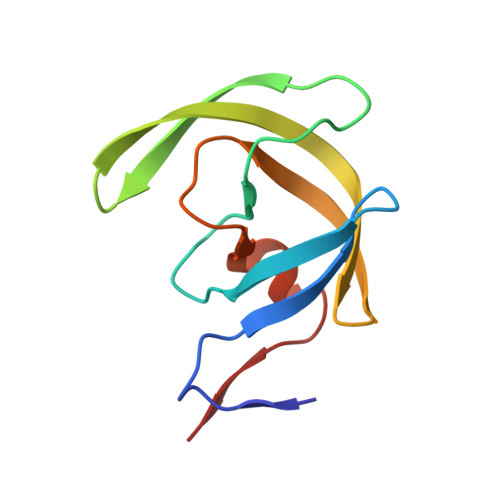
4DJQ

Crystal Structure of wild-type HIV-1 Protease in Complex with MKP86


Experimental Data Snapshot

  • Method: X-RAY DIFFRACTION
  • Resolution: 1.40 Å
  • R-Value Free: 0.204 
  • R-Value Work: 0.179 
  • R-Value Observed: 0.180 

wwPDB Validation   3D Report Full Report


Ligand Structure Quality Assessment 


This is version 1.3 of the entry. See complete history


Literature

Design, synthesis, and biological and structural evaluations of novel HIV-1 protease inhibitors to combat drug resistance.

Parai, M.K.Huggins, D.J.Cao, H.Nalam, M.N.Ali, A.Schiffer, C.A.Tidor, B.Rana, T.M.

(2012) J Med Chem 55: 6328-6341

  • DOI: https://doi.org/10.1021/jm300238h
  • Primary Citation of Related Structures:  
    4DJO, 4DJP, 4DJQ, 4DJR

  • PubMed Abstract: 

    A series of new HIV-1 protease inhibitors (PIs) were designed using a general strategy that combines computational structure-based design with substrate-envelope constraints. The PIs incorporate various alcohol-derived P2 carbamates with acyclic and cyclic heteroatomic functionalities into the (R)-hydroxyethylamine isostere. Most of the new PIs show potent binding affinities against wild-type HIV-1 protease and three multidrug resistant (MDR) variants. In particular, inhibitors containing the 2,2-dichloroacetamide, pyrrolidinone, imidazolidinone, and oxazolidinone moieties at P2 are the most potent with K(i) values in the picomolar range. Several new PIs exhibit nanomolar antiviral potencies against patient-derived wild-type viruses from HIV-1 clades A, B, and C and two MDR variants. Crystal structure analyses of four potent inhibitors revealed that carbonyl groups of the new P2 moieties promote extensive hydrogen bond interactions with the invariant Asp29 residue of the protease. These structure-activity relationship findings can be utilized to design new PIs with enhanced enzyme inhibitory and antiviral potencies.


  • Organizational Affiliation

    Program for RNA Biology, Sanford-Burnham Medical Research Institute, La Jolla, CA 92037, USA.


Macromolecules
Find similar proteins by:  (by identity cutoff)  |  3D Structure
Entity ID: 1
MoleculeChains Sequence LengthOrganismDetailsImage
Pol polyprotein
A, B
99Human immunodeficiency virus 1Mutation(s): 1 
Gene Names: gag-polpol
UniProt
Find proteins for Q90K99 (Human immunodeficiency virus 1)
Explore Q90K99 
Go to UniProtKB:  Q90K99
Entity Groups  
Sequence Clusters30% Identity50% Identity70% Identity90% Identity95% Identity100% Identity
UniProt GroupQ90K99
Sequence Annotations
Expand
  • Reference Sequence
Small Molecules
Ligands 2 Unique
IDChains Name / Formula / InChI Key2D Diagram3D Interactions
M86
Query on M86

Download Ideal Coordinates CCD File 
C [auth A]2-(2-oxoimidazolidin-1-yl)ethyl [(2S,3R)-3-hydroxy-4-{[(4-methoxyphenyl)sulfonyl][(2S)-2-methylbutyl]amino}-1-phenylbutan-2-yl]carbamate
C28 H40 N4 O7 S
KVKPWRDMMABYRK-OUIFVKKZSA-N
PO4
Query on PO4

Download Ideal Coordinates CCD File 
D [auth B],
E [auth B]
PHOSPHATE ION
O4 P
NBIIXXVUZAFLBC-UHFFFAOYSA-K
Binding Affinity Annotations 
IDSourceBinding Affinity
M86 PDBBind:  4DJQ Ki: 0.05 (nM) from 1 assay(s)
Experimental Data & Validation

Experimental Data

  • Method: X-RAY DIFFRACTION
  • Resolution: 1.40 Å
  • R-Value Free: 0.204 
  • R-Value Work: 0.179 
  • R-Value Observed: 0.180 
  • Space Group: P 21 21 21
Unit Cell:
Length ( Å )Angle ( ˚ )
a = 50.711α = 90
b = 57.93β = 90
c = 61.815γ = 90
Software Package:
Software NamePurpose
DENZOdata reduction
SCALEPACKdata scaling
REFMACrefinement
PDB_EXTRACTdata extraction
HKL-2000data collection
HKL-2000data reduction
AMoREphasing

Structure Validation

View Full Validation Report


Ligand Structure Quality Assessment 


Entry History 

Deposition Data

Revision History  (Full details and data files)

  • Version 1.0: 2012-08-01
    Type: Initial release
  • Version 1.1: 2013-01-02
    Changes: Database references
  • Version 1.2: 2017-11-15
    Changes: Refinement description
  • Version 1.3: 2024-02-28
    Changes: Data collection, Database references, Derived calculations