


Identification of Novel Peptide Deformylase Inhibitors from Natural Products
Cui, K., Zhu, L., Lu, W., Huang, J.To be published.
Experimental Data Snapshot
Starting Model: experimental
View more details
Entity ID: 1 | |||||
|---|---|---|---|---|---|
| Molecule | Chains | Sequence Length | Organism | Details | Image |
| Peptide deformylase 11 | 181 | Helicobacter pylori | Mutation(s): 0 Gene Names: def, def11 EC: 3.5.1.88 | 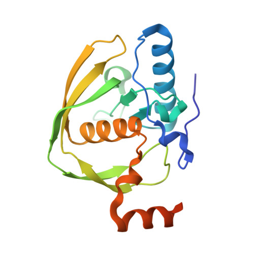 | |
UniProt | |||||
Entity Groups | |||||
| Sequence Clusters | 30% Identity50% Identity70% Identity90% Identity95% Identity100% Identity | ||||
| UniProt Group | Q672W7 | ||||
Sequence AnnotationsExpand | |||||
Reference Sequence | |||||
| Ligands 4 Unique | |||||
|---|---|---|---|---|---|
| ID | Chains | Name / Formula / InChI Key | 2D Diagram | 3D Interactions | |
| QAP Download:Ideal Coordinates CCD File | C [auth A] | 2-phenylethyl (2E)-3-(3,4-dihydroxyphenyl)prop-2-enoate C17 H16 O4 SWUARLUWKZWEBQ-VQHVLOKHSA-N |  | ||
| EPE Download:Ideal Coordinates CCD File | D [auth A] | 4-(2-HYDROXYETHYL)-1-PIPERAZINE ETHANESULFONIC ACID C8 H18 N2 O4 S JKMHFZQWWAIEOD-UHFFFAOYSA-N |  | ||
| DMS Download:Ideal Coordinates CCD File | E [auth A], F [auth A] | DIMETHYL SULFOXIDE C2 H6 O S IAZDPXIOMUYVGZ-UHFFFAOYSA-N |  | ||
| CO Download:Ideal Coordinates CCD File | B [auth A] | COBALT (II) ION Co XLJKHNWPARRRJB-UHFFFAOYSA-N |  | ||
| Length ( Å ) | Angle ( ˚ ) |
|---|---|
| a = 41.891 | α = 90 |
| b = 52.057 | β = 90 |
| c = 92.064 | γ = 90 |
| Software Name | Purpose |
|---|---|
| HKL-2000 | data collection |
| PHENIX | refinement |
| HKL-2000 | data reduction |
| HKL-2000 | data scaling |


