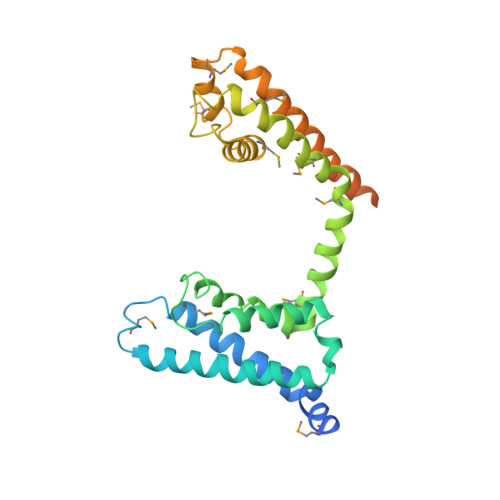
4EKW

Crystal structure of the NavAb voltage-gated sodium channel (wild-type, 3.2 A)


Experimental Data Snapshot

  • Method: X-RAY DIFFRACTION
  • Resolution: 3.21 Å
  • R-Value Free: 0.322 
  • R-Value Work: 0.308 
  • R-Value Observed: 0.309 

Starting Model: experimental
View more details

wwPDB Validation   3D Report Full Report


Ligand Structure Quality Assessment 


This is version 1.4 of the entry. See complete history


Literature

Crystal structure of a voltage-gated sodium channel in two potentially inactivated states.

Payandeh, J.Gamal El-Din, T.M.Scheuer, T.Zheng, N.Catterall, W.A.

(2012) Nature 486: 135-139

  • DOI: https://doi.org/10.1038/nature11077
  • Primary Citation of Related Structures:  
    4EKW

  • PubMed Abstract: 

    In excitable cells, voltage-gated sodium (Na(V)) channels activate to initiate action potentials and then undergo fast and slow inactivation processes that terminate their ionic conductance. Inactivation is a hallmark of Na(V) channel function and is critical for control of membrane excitability, but the structural basis for this process has remained elusive. Here we report crystallographic snapshots of the wild-type Na(V)Ab channel from Arcobacter butzleri captured in two potentially inactivated states at 3.2 Å resolution. Compared to previous structures of Na(V)Ab channels with cysteine mutations in the pore-lining S6 helices (ref. 4), the S6 helices and the intracellular activation gate have undergone significant rearrangements: one pair of S6 helices has collapsed towards the central pore axis and the other S6 pair has moved outward to produce a striking dimer-of-dimers configuration. An increase in global structural asymmetry is observed throughout our wild-type Na(V)Ab models, reshaping the ion selectivity filter at the extracellular end of the pore, the central cavity and its residues that are analogous to the mammalian drug receptor site, and the lateral pore fenestrations. The voltage-sensing domains have also shifted around the perimeter of the pore module in wild-type Na(V)Ab, compared to the mutant channel, and local structural changes identify a conserved interaction network that connects distant molecular determinants involved in Na(V) channel gating and inactivation. These potential inactivated-state structures provide new insights into Na(V) channel gating and novel avenues to drug development and therapy for a range of debilitating Na(V) channelopathies.


  • Organizational Affiliation

    Department of Pharmacology, University of Washington, Seattle, Washington 98195, USA.


Macromolecules
Find similar proteins by:  (by identity cutoff)  |  3D Structure
Entity ID: 1
MoleculeChains Sequence LengthOrganismDetailsImage
Ion transport protein
A, B, C, D
285Aliarcobacter butzleri RM4018Mutation(s): 0 
Gene Names: Abu_1752
Membrane Entity: Yes 
UniProt
Find proteins for A8EVM5 (Aliarcobacter butzleri (strain RM4018))
Explore A8EVM5 
Go to UniProtKB:  A8EVM5
Entity Groups  
Sequence Clusters30% Identity50% Identity70% Identity90% Identity95% Identity100% Identity
UniProt GroupA8EVM5
Sequence Annotations
Expand
  • Reference Sequence
Small Molecules
Ligands 2 Unique
IDChains Name / Formula / InChI Key2D Diagram3D Interactions
PX4
Query on PX4

Download Ideal Coordinates CCD File 
E [auth A],
G [auth B],
H [auth B],
J [auth C],
L [auth D]
1,2-DIMYRISTOYL-SN-GLYCERO-3-PHOSPHOCHOLINE
C36 H73 N O8 P
CITHEXJVPOWHKC-UUWRZZSWSA-O
PO4
Query on PO4

Download Ideal Coordinates CCD File 
F [auth A],
I [auth B],
K [auth C],
M [auth D]
PHOSPHATE ION
O4 P
NBIIXXVUZAFLBC-UHFFFAOYSA-K
Modified Residues  1 Unique
IDChains TypeFormula2D DiagramParent
MSE
Query on MSE
A, B, C, D
L-PEPTIDE LINKINGC5 H11 N O2 SeMET
Experimental Data & Validation

Experimental Data

  • Method: X-RAY DIFFRACTION
  • Resolution: 3.21 Å
  • R-Value Free: 0.322 
  • R-Value Work: 0.308 
  • R-Value Observed: 0.309 
  • Space Group: P 42
Unit Cell:
Length ( Å )Angle ( ˚ )
a = 125.856α = 90
b = 125.856β = 90
c = 192.641γ = 90
Software Package:
Software NamePurpose
HKL-2000data collection
PHENIXmodel building
REFMACrefinement
DENZOdata reduction
SCALEPACKdata scaling
PHENIXphasing

Structure Validation

View Full Validation Report


Ligand Structure Quality Assessment 


Entry History 

Deposition Data

Revision History  (Full details and data files)

  • Version 1.0: 2012-05-16
    Type: Initial release
  • Version 1.1: 2012-10-03
    Changes: Database references
  • Version 1.2: 2023-09-13
    Changes: Data collection, Database references, Derived calculations, Refinement description
  • Version 1.3: 2023-12-06
    Changes: Data collection
  • Version 1.4: 2024-10-16
    Changes: Structure summary