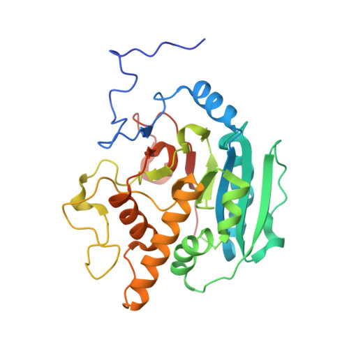
pH-induced conformational changes in human ABO(H) blood group glycosyltransferases confirm the importance of electrostatic interactions in the formation of the semi-closed state.
Johal, A.R., Blackler, R.J., Alfaro, J.A., Schuman, B., Borisova, S., Evans, S.V.(2014) Glycobiology 24: 237-246
- PubMed: 24265507
- DOI: https://doi.org/10.1093/glycob/cwt098
- Primary Citation of Related Structures:
4FQW, 4FRA, 4FRB, 4FRD, 4FRE, 4FRH, 4FRL, 4FRM, 4FRO, 4FRP, 4FRQ, 4GBP, 4KXO - PubMed Abstract:
The homologous human ABO(H) A and B blood group glycosyltransferases GTA and GTB have two mobile polypeptide loops surrounding their active sites that serve to allow substrate access and product egress and to recognize and sequester substrates for catalysis. Previous studies have established that these enzymes can move from the "open" state to the "semi-closed" then "closed" states in response to addition of a substrate. The contribution of electrostatic interactions to these conformational changes has now been demonstrated by the determination at various pH of the structures of GTA, GTB and the chimeric enzyme ABBA. At near-neutral pH, GTA displays the closed state in which both mobile loops order around the active site, whereas ABBA and GTB display the open state. At low pH, the apparent protonation of the DXD motif in GTA leads to the expulsion of the donor analog to yield the open state, whereas at high pH, both ABBA and GTB form the semi-closed state in which the first mobile loop becomes an ordered α-helix. Step-wise deprotonation of GTB in increments of 0.5 between pH 6.5 and 10.0 shows that helix ordering is gradual, which indicates that the formation of the semi-closed state is dependent on electrostatic forces consistent with the binding of substrate. Spectropolarimetric studies of the corresponding stand-alone peptide in solution reveal no tendency toward helix formation from pH 7.0 to 10.0, which shows that pH-dependent stability is a product of the larger protein environment and underlines the importance of substrate in active site ordering.
Organizational Affiliation:
Department of Biochemistry and Microbiology, University of Victoria, Victoria, BC, Canada V8W 3P6.


