4G1M

Re-refinement of alpha V beta 3 structure


Experimental Data Snapshot

  • Method: X-RAY DIFFRACTION
  • Resolution: 2.90 Å
  • R-Value Free: 
    0.233 (Depositor), 0.241 (DCC) 
  • R-Value Work: 
    0.179 (Depositor), 0.186 (DCC) 
  • R-Value Observed: 
    0.182 (Depositor) 

wwPDB Validation   3D Report Full Report


Ligand Structure Quality Assessment 

Created with Raphaël 2.3.0Worse 01 BetterLigand structure goodness of fit to experimental dataBest fitted NAGClick on this verticalbar to view details

This is version 2.1 of the entry. See complete history


Literature

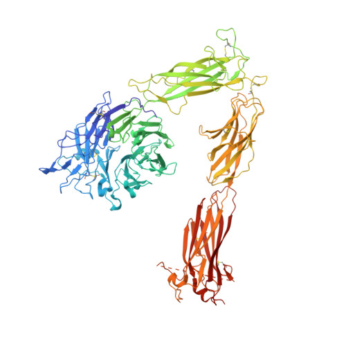
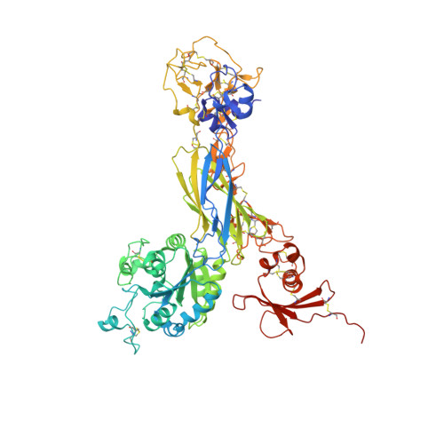
Alpha V Beta 3 Integrin Crystal Structures and their Functional Implications

Dong, X.Mi, L.Z.Zhu, J.Wang, W.Hu, P.Luo, B.H.Springer, T.A.

(2012) Biochemistry 51: 8814-8828

  • DOI: https://doi.org/10.1021/bi300734n
  • Primary Citation of Related Structures:  
    4G1E, 4G1M

  • PubMed Abstract: 

    Many questions about the significance of structural features of integrin α(V)β(3) with respect to its mechanism of activation remain. We have determined and re-refined crystal structures of the α(V)β(3) ectodomain linked to C-terminal coiled coils (α(V)β(3)-AB) and four transmembrane (TM) residues in each subunit (α(V)β(3)-1TM), respectively. The α(V) and β(3) subunits with four and eight extracellular domains, respectively, are bent at knees between the integrin headpiece and lower legs, and the headpiece has the closed, low-affinity conformation. The structures differ in the occupancy of three metal-binding sites in the βI domain. Occupancy appears to be related to the pH of crystallization, rather than to the physiologic regulation of ligand binding at the central, metal ion-dependent adhesion site. No electron density was observed for TM residues and much of the α(V) linker. α(V)β(3)-AB and α(V)β(3)-1TM demonstrate flexibility in the linker between their extracellular and TM domains, rather than the previously proposed rigid linkage. A previously postulated interface between the α(V) and β(3) subunits at their knees was also not supported, because it lacks high-quality density, required rebuilding in α(V)β(3)-1TM, and differed markedly between α(V)β(3)-1TM and α(V)β(3)-AB. Together with the variation in domain-domain orientation within their bent ectodomains between α(V)β(3)-AB and α(V)β(3)-1TM, these findings are compatible with the requirement for large structural changes, such as extension at the knees and headpiece opening, in conveying activation signals between the extracellular ligand-binding site and the cytoplasm.


  • Organizational Affiliation

    Immune Disease Institute, Children's Hospital Boston, and Department of Biological Chemistry and Molecular Pharmacology, Harvard Medical School, 3 Blackfan Circle, Boston, Massachusetts 02115, United States.


Macromolecules
Find similar proteins by:  (by identity cutoff)  |  3D Structure
Entity ID: 1
MoleculeChains Sequence LengthOrganismDetailsImage
Integrin alpha-V959Homo sapiensMutation(s): 0 
Gene Names: ITGAVMSK8VNRA
UniProt & NIH Common Fund Data Resources
Find proteins for P06756 (Homo sapiens)
Explore P06756 
Go to UniProtKB:  P06756
PHAROS:  P06756
GTEx:  ENSG00000138448 
Entity Groups  
Sequence Clusters30% Identity50% Identity70% Identity90% Identity95% Identity100% Identity
UniProt GroupP06756
Glycosylation
Glycosylation Sites: 11Go to GlyGen: P06756-1
Sequence Annotations
Expand
  • Reference Sequence
Find similar proteins by:  (by identity cutoff)  |  3D Structure
Entity ID: 2
MoleculeChains Sequence LengthOrganismDetailsImage
Integrin beta-3692Homo sapiensMutation(s): 0 
Gene Names: ITGB3GP3A
UniProt & NIH Common Fund Data Resources
Find proteins for P05106 (Homo sapiens)
Explore P05106 
Go to UniProtKB:  P05106
PHAROS:  P05106
GTEx:  ENSG00000259207 
Entity Groups  
Sequence Clusters30% Identity50% Identity70% Identity90% Identity95% Identity100% Identity
UniProt GroupP05106
Glycosylation
Glycosylation Sites: 5Go to GlyGen: P05106-1
Sequence Annotations
Expand
  • Reference Sequence
Oligosaccharides

Help

Entity ID: 3
MoleculeChains Length2D Diagram Glycosylation3D Interactions
alpha-D-mannopyranose-(1-3)-[alpha-D-mannopyranose-(1-6)]beta-D-mannopyranose-(1-4)-2-acetamido-2-deoxy-beta-D-glucopyranose-(1-4)-2-acetamido-2-deoxy-beta-D-glucopyranose
C
5N-Glycosylation
Glycosylation Resources
GlyTouCan:  G22768VO
GlyCosmos:  G22768VO
GlyGen:  G22768VO
Entity ID: 4
MoleculeChains Length2D Diagram Glycosylation3D Interactions
2-acetamido-2-deoxy-beta-D-glucopyranose-(1-4)-2-acetamido-2-deoxy-beta-D-glucopyranose
D, F, G, H, I
2N-Glycosylation
Glycosylation Resources
GlyTouCan:  G42666HT
GlyCosmos:  G42666HT
GlyGen:  G42666HT
Entity ID: 5
MoleculeChains Length2D Diagram Glycosylation3D Interactions
alpha-D-mannopyranose-(1-2)-alpha-D-mannopyranose-(1-3)-[alpha-D-mannopyranose-(1-3)-alpha-D-mannopyranose-(1-6)]beta-D-mannopyranose-(1-4)-2-acetamido-2-deoxy-beta-D-glucopyranose-(1-4)-2-acetamido-2-deoxy-beta-D-glucopyranose
E
7N-Glycosylation
Glycosylation Resources
GlyTouCan:  G07617FP
GlyCosmos:  G07617FP
GlyGen:  G07617FP
Entity ID: 6
MoleculeChains Length2D Diagram Glycosylation3D Interactions
beta-D-mannopyranose-(1-4)-2-acetamido-2-deoxy-beta-D-glucopyranose-(1-4)-2-acetamido-2-deoxy-beta-D-glucopyranose
N
3N-Glycosylation
Glycosylation Resources
GlyTouCan:  G15407YE
GlyCosmos:  G15407YE
GlyGen:  G15407YE
Small Molecules
Ligands 3 Unique
IDChains Name / Formula / InChI Key2D Diagram3D Interactions
NAG
Query on NAG

Download Ideal Coordinates CCD File 
T [auth A],
U [auth A],
Y [auth B],
Z [auth B]
2-acetamido-2-deoxy-beta-D-glucopyranose
C8 H15 N O6
OVRNDRQMDRJTHS-FMDGEEDCSA-N
CA
Query on CA

Download Ideal Coordinates CCD File 
O [auth A]
P [auth A]
Q [auth A]
R [auth A]
S [auth A]
CALCIUM ION
Ca
BHPQYMZQTOCNFJ-UHFFFAOYSA-N
NA
Query on NA

Download Ideal Coordinates CCD File 
V [auth A]SODIUM ION
Na
FKNQFGJONOIPTF-UHFFFAOYSA-N
Experimental Data & Validation

Experimental Data

  • Method: X-RAY DIFFRACTION
  • Resolution: 2.90 Å
  • R-Value Free:  0.233 (Depositor), 0.241 (DCC) 
  • R-Value Work:  0.179 (Depositor), 0.186 (DCC) 
  • R-Value Observed: 0.182 (Depositor) 
Space Group: P 32 2 1
Unit Cell:
Length ( Å )Angle ( ˚ )
a = 129.87α = 90
b = 129.87β = 90
c = 305.9γ = 120
Software Package:
Software NamePurpose
PHENIXrefinement
CNSrefinement
HKL-2000data collection
DENZOdata reduction
HKL-2000data scaling
CNSphasing

Structure Validation

View Full Validation Report


Ligand Structure Quality Assessment 

Created with Raphaël 2.3.0Worse 01 BetterLigand structure goodness of fit to experimental dataBest fitted NAGClick on this verticalbar to view details

Entry History 

Deposition Data

Revision History  (Full details and data files)

  • Version 1.0: 2012-12-12
    Type: Initial release
  • Version 1.1: 2012-12-19
    Changes: Database references
  • Version 1.2: 2019-08-14
    Changes: Data collection, Derived calculations, Refinement description
  • Version 2.0: 2020-07-29
    Type: Remediation
    Reason: Carbohydrate remediation
    Changes: Advisory, Atomic model, Data collection, Derived calculations, Structure summary
  • Version 2.1: 2024-11-06
    Changes: Data collection, Database references, Derived calculations, Structure summary