4G74

Crystal structure of NDH with Quinone


Experimental Data Snapshot

  • Method: X-RAY DIFFRACTION
  • Resolution: 2.48 Å
  • R-Value Free: 
    0.233 (Depositor), 0.230 (DCC) 
  • R-Value Work: 
    0.195 (Depositor), 0.190 (DCC) 
  • R-Value Observed: 
    0.197 (Depositor) 

Starting Model: experimental
View more details

wwPDB Validation   3D Report Full Report


Ligand Structure Quality Assessment 

Created with Raphaël 2.3.0Worse 01 BetterLigand structure goodness of fit to experimental dataBest fitted FADClick on this verticalbar to view detailsBest fitted UQ5Click on this verticalbar to view detailsBest fitted TRTClick on this verticalbar to view details

This is version 1.2 of the entry. See complete history


Literature

Structural insight into the type-II mitochondrial NADH dehydrogenases.

Feng, Y.Li, W.Li, J.Wang, J.Ge, J.Xu, D.Liu, Y.Wu, K.Zeng, Q.Wu, J.W.Tian, C.Zhou, B.Yang, M.

(2012) Nature 491: 478-482

  • DOI: https://doi.org/10.1038/nature11541
  • Primary Citation of Related Structures:  
    4G6G, 4G6H, 4G73, 4G74

  • PubMed Abstract: 

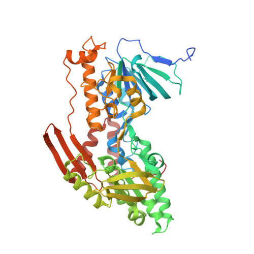
    The single-component type-II NADH dehydrogenases (NDH-2s) serve as alternatives to the multisubunit respiratory complex I (type-I NADH dehydrogenase (NDH-1), also called NADH:ubiquinone oxidoreductase; EC 1.6.5.3) in catalysing electron transfer from NADH to ubiquinone in the mitochondrial respiratory chain. The yeast NDH-2 (Ndi1) oxidizes NADH on the matrix side and reduces ubiquinone to maintain mitochondrial NADH/NAD(+) homeostasis. Ndi1 is a potential therapeutic agent for human diseases caused by complex I defects, particularly Parkinson's disease, because its expression restores the mitochondrial activity in animals with complex I deficiency. NDH-2s in pathogenic microorganisms are viable targets for new antibiotics. Here we solve the crystal structures of Ndi1 in its substrate-free, NADH-, ubiquinone- and NADH-ubiquinone-bound states, to help understand the catalytic mechanism of NDH-2s. We find that Ndi1 homodimerization through its carboxy-terminal domain is critical for its catalytic activity and membrane targeting. The structures reveal two ubiquinone-binding sites (UQ(I) and UQ(II)) in Ndi1. NADH and UQ(I) can bind to Ndi1 simultaneously to form a substrate-protein complex. We propose that UQ(I) interacts with FAD to act as an intermediate for electron transfer, and that NADH transfers electrons through this FAD-UQ(I) complex to UQ(II). Together our data reveal the regulatory and catalytic mechanisms of Ndi1 and may facilitate the development or targeting of NDH-2s for potential therapeutic applications.


  • Organizational Affiliation

    State Key Laboratory of Biomembrane and Membrane Biotechnology, Tsinghua-Peking Center for Life Sciences, School of Life Sciences, Tsinghua University, Beijing 100084, China.


Macromolecules
Find similar proteins by:  (by identity cutoff)  |  3D Structure
Entity ID: 1
MoleculeChains Sequence LengthOrganismDetailsImage
Rotenone-insensitive NADH-ubiquinone oxidoreductase, mitochondrial
A, B
502Saccharomyces cerevisiae S288CMutation(s): 0 
Gene Names: NDI1YML120CYM7056.06C
EC: 1.6.5.9
Membrane Entity: Yes 
UniProt
Find proteins for P32340 (Saccharomyces cerevisiae (strain ATCC 204508 / S288c))
Explore P32340 
Go to UniProtKB:  P32340
Entity Groups  
Sequence Clusters30% Identity50% Identity70% Identity90% Identity95% Identity100% Identity
UniProt GroupP32340
Sequence Annotations
Expand
  • Reference Sequence
Small Molecules
Ligands 4 Unique
IDChains Name / Formula / InChI Key2D Diagram3D Interactions
FAD
Query on FAD

Download Ideal Coordinates CCD File 
I [auth A],
S [auth B]
FLAVIN-ADENINE DINUCLEOTIDE
C27 H33 N9 O15 P2
VWWQXMAJTJZDQX-UYBVJOGSSA-N
UQ5
Query on UQ5

Download Ideal Coordinates CCD File 
C [auth A],
D [auth A],
O [auth B],
P [auth B]
2,3-DIMETHOXY-5-METHYL-6-(3,11,15,19-TETRAMETHYL-EICOSA-2,6,10,14,18-PENTAENYL)-[1,4]BENZOQUINONE
C34 H50 O4
NYFAQDMDAFCWPU-UVCHAVPFSA-N
TRT
Query on TRT

Download Ideal Coordinates CCD File 
E [auth A],
F [auth A],
G [auth A],
Q [auth B]
FRAGMENT OF TRITON X-100
C21 H36 O4
HEUDUECKTWTQQR-UHFFFAOYSA-N
MG
Query on MG

Download Ideal Coordinates CCD File 
H [auth A]
J [auth A]
K [auth A]
L [auth A]
M [auth A]
MAGNESIUM ION
Mg
JLVVSXFLKOJNIY-UHFFFAOYSA-N
Experimental Data & Validation

Experimental Data

  • Method: X-RAY DIFFRACTION
  • Resolution: 2.48 Å
  • R-Value Free:  0.233 (Depositor), 0.230 (DCC) 
  • R-Value Work:  0.195 (Depositor), 0.190 (DCC) 
  • R-Value Observed: 0.197 (Depositor) 
Space Group: C 2 2 21
Unit Cell:
Length ( Å )Angle ( ˚ )
a = 132.909α = 90
b = 230.844β = 90
c = 112.45γ = 90
Software Package:
Software NamePurpose
HKL-2000data collection
PHASESphasing
PHENIXrefinement
HKL-2000data reduction
HKL-2000data scaling

Structure Validation

View Full Validation Report


Ligand Structure Quality Assessment 

Created with Raphaël 2.3.0Worse 01 BetterLigand structure goodness of fit to experimental dataBest fitted FADClick on this verticalbar to view detailsBest fitted UQ5Click on this verticalbar to view detailsBest fitted TRTClick on this verticalbar to view details

Entry History 

Deposition Data

Revision History  (Full details and data files)

  • Version 1.0: 2012-10-24
    Type: Initial release
  • Version 1.1: 2012-12-05
    Changes: Database references
  • Version 1.2: 2023-11-08
    Changes: Data collection, Database references, Derived calculations, Refinement description