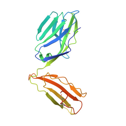
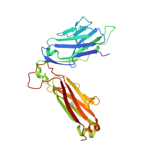
4G8F

Crystal Structure of clone42 TCR


Experimental Data Snapshot

  • Method: X-RAY DIFFRACTION
  • Resolution: 2.10 Å
  • R-Value Free: 0.236 
  • R-Value Work: 0.203 
  • R-Value Observed: 0.204 

Starting Model: experimental
View more details

wwPDB Validation   3D Report Full Report


This is version 1.4 of the entry. See complete history


Literature

A conserved human T cell population targets mycobacterial antigens presented by CD1b.

Van Rhijn, I.Kasmar, A.de Jong, A.Gras, S.Bhati, M.Doorenspleet, M.E.de Vries, N.Godfrey, D.I.Altman, J.D.de Jager, W.Rossjohn, J.Moody, D.B.

(2013) Nat Immunol 14: 706-713

  • DOI: https://doi.org/10.1038/ni.2630
  • Primary Citation of Related Structures:  
    4G8E, 4G8F

  • PubMed Abstract: 

    Human T cell antigen receptors (TCRs) pair in millions of combinations to create complex and unique T cell repertoires for each person. Through the use of tetramers to analyze TCRs reactive to the antigen-presenting molecule CD1b, we detected T cells with highly stereotyped TCR α-chains present among genetically unrelated patients with tuberculosis. The germline-encoded, mycolyl lipid-reactive (GEM) TCRs had an α-chain bearing the variable (V) region TRAV1-2 rearranged to the joining (J) region TRAJ9 with few nontemplated (N)-region additions. Analysis of TCRs by high-throughput sequencing, binding and crystallography showed linkage of TCRα sequence motifs to high-affinity recognition of antigen. Thus, the CD1-reactive TCR repertoire is composed of at least two compartments: high-affinity GEM TCRs, and more-diverse TCRs with low affinity for CD1b-lipid complexes. We found high interdonor conservation of TCRs that probably resulted from selection by a nonpolymorphic antigen-presenting molecule and an immunodominant antigen.


  • Organizational Affiliation

    Division of Rheumatology, Immunology and Allergy, Brigham and Women's Hospital, Harvard Medical School, Boston, Massachusetts, USA. i.vanrhijn@uu.nl


Macromolecules
Find similar proteins by:  (by identity cutoff)  |  3D Structure
Entity ID: 1
MoleculeChains Sequence LengthOrganismDetailsImage
alpha chain clone 42 TCR204Homo sapiensMutation(s): 0 
UniProt & NIH Common Fund Data Resources
Find proteins for P01848 (Homo sapiens)
Explore P01848 
Go to UniProtKB:  P01848
PHAROS:  P01848
Entity Groups  
Sequence Clusters30% Identity50% Identity70% Identity90% Identity95% Identity100% Identity
UniProt GroupP01848
Sequence Annotations
Expand
  • Reference Sequence
Find similar proteins by:  (by identity cutoff)  |  3D Structure
Entity ID: 2
MoleculeChains Sequence LengthOrganismDetailsImage
beta chain clone 42 TCR243Homo sapiensMutation(s): 0 
UniProt
Find proteins for P01850 (Homo sapiens)
Explore P01850 
Go to UniProtKB:  P01850
Entity Groups  
Sequence Clusters30% Identity50% Identity70% Identity90% Identity95% Identity100% Identity
UniProt GroupP01850
Sequence Annotations
Expand
  • Reference Sequence
Experimental Data & Validation

Experimental Data

  • Method: X-RAY DIFFRACTION
  • Resolution: 2.10 Å
  • R-Value Free: 0.236 
  • R-Value Work: 0.203 
  • R-Value Observed: 0.204 
  • Space Group: P 1 21 1
Unit Cell:
Length ( Å )Angle ( ˚ )
a = 46.52α = 90
b = 56.79β = 101.43
c = 85.89γ = 90
Software Package:
Software NamePurpose
XSCALEdata scaling
PHASERphasing
BUSTER-TNTrefinement
PDB_EXTRACTdata extraction
Blu-Icedata collection
XDSdata reduction
BUSTERrefinement

Structure Validation

View Full Validation Report


Entry History 

Deposition Data

Revision History  (Full details and data files)

  • Version 1.0: 2013-05-08
    Type: Initial release
  • Version 1.1: 2013-07-03
    Changes: Database references
  • Version 1.2: 2013-07-10
    Changes: Database references
  • Version 1.3: 2017-10-11
    Changes: Data collection, Refinement description
  • Version 1.4: 2023-09-13
    Changes: Data collection, Database references, Refinement description