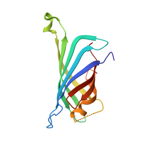
4GDA | pdb_00004gda

Circular Permuted Streptavidin A50/N49


Experimental Data Snapshot

  • Method: X-RAY DIFFRACTION
  • Resolution: 1.00 Å
  • R-Value Free: 
    0.143 (Depositor), 0.150 (DCC) 
  • R-Value Work: 
    0.118 (Depositor), 0.130 (DCC) 
  • R-Value Observed: 
    0.119 (Depositor) 

wwPDB Validation   3D Report Full Report


Ligand Structure Quality Assessment 

Created with Raphaël 2.3.0Worse 01 BetterLigand structure goodness of fit to experimental dataBest fitted BTNClick on this verticalbar to view details

This is version 1.3 of the entry. See complete history


Literature

Structural consequences of cutting a binding loop: two circularly permuted variants of streptavidin.

Le Trong, I.Chu, V.Xing, Y.Lybrand, T.P.Stayton, P.S.Stenkamp, R.E.

(2013) Acta Crystallogr D Biol Crystallogr 69: 968-977

  • DOI: https://doi.org/10.1107/S0907444913003855
  • Primary Citation of Related Structures:  
    4GD9, 4GDA

  • PubMed Abstract: 

    Circular permutation of streptavidin was carried out in order to investigate the role of a main-chain amide in stabilizing the high-affinity complex of the protein and biotin. Mutant proteins CP49/48 and CP50/49 were constructed to place new N-termini at residues 49 and 50 in a flexible loop involved in stabilizing the biotin complex. Crystal structures of the two mutants show that half of each loop closes over the binding site, as observed in wild-type streptavidin, while the other half adopts the open conformation found in the unliganded state. The structures are consistent with kinetic and thermodynamic data and indicate that the loop plays a role in enthalpic stabilization of the bound state via the Asn49 amide-biotin hydrogen bond. In wild-type streptavidin, the entropic penalties of immobilizing a flexible portion of the protein to enhance binding are kept to a manageable level by using a contiguous loop of medium length (six residues) which is already constrained by its anchorage to strands of the β-barrel protein. A molecular-dynamics simulation for CP50/49 shows that cleavage of the binding loop results in increased structural fluctuations for Ser45 and that these fluctuations destabilize the streptavidin-biotin complex.


  • Organizational Affiliation

    Department of Biological Structure, University of Washington, Box 357420, Seattle, WA 98195-7420, USA.


Macromolecules
Find similar proteins by:  (by identity cutoff)  |  3D Structure
Entity ID: 1
MoleculeChains Sequence LengthOrganismDetailsImage
Streptavidin
A, B
131Streptomyces avidiniiMutation(s): 0 
UniProt
Find proteins for P22629 (Streptomyces avidinii)
Explore P22629 
Go to UniProtKB:  P22629
Entity Groups  
Sequence Clusters30% Identity50% Identity70% Identity90% Identity95% Identity100% Identity
UniProt GroupP22629
Sequence Annotations
Expand
  • Reference Sequence
Small Molecules
Ligands 4 Unique
IDChains Name / Formula / InChI Key2D Diagram3D Interactions
BTN
Query on BTN

Download Ideal Coordinates CCD File 
C [auth A],
G [auth B]
BIOTIN
C10 H16 N2 O3 S
YBJHBAHKTGYVGT-ZKWXMUAHSA-N
SO4
Query on SO4

Download Ideal Coordinates CCD File 
F [auth A],
I [auth B],
J [auth B]
SULFATE ION
O4 S
QAOWNCQODCNURD-UHFFFAOYSA-L
GOL
Query on GOL

Download Ideal Coordinates CCD File 
D [auth A],
E [auth A],
H [auth B]
GLYCEROL
C3 H8 O3
PEDCQBHIVMGVHV-UHFFFAOYSA-N
EOH
Query on EOH

Download Ideal Coordinates CCD File 
K [auth B]ETHANOL
C2 H6 O
LFQSCWFLJHTTHZ-UHFFFAOYSA-N
Experimental Data & Validation

Experimental Data

  • Method: X-RAY DIFFRACTION
  • Resolution: 1.00 Å
  • R-Value Free:  0.143 (Depositor), 0.150 (DCC) 
  • R-Value Work:  0.118 (Depositor), 0.130 (DCC) 
  • R-Value Observed: 0.119 (Depositor) 
Space Group: I 2 2 2
Unit Cell:
Length ( Å )Angle ( ˚ )
a = 46.809α = 90
b = 94.027β = 90
c = 104.279γ = 90
Software Package:
Software NamePurpose
SCALEPACKdata scaling
REFMACrefinement
PDB_EXTRACTdata extraction
Blu-Icedata collection
DENZOdata reduction

Structure Validation

View Full Validation Report


Ligand Structure Quality Assessment 

Created with Raphaël 2.3.0Worse 01 BetterLigand structure goodness of fit to experimental dataBest fitted BTNClick on this verticalbar to view details

Entry History 

Deposition Data

Revision History  (Full details and data files)

  • Version 1.0: 2013-06-05
    Type: Initial release
  • Version 1.1: 2017-08-02
    Changes: Refinement description, Source and taxonomy
  • Version 1.2: 2017-11-15
    Changes: Refinement description, Source and taxonomy
  • Version 1.3: 2024-02-28
    Changes: Data collection, Database references, Derived calculations