4GLV | pdb_00004glv

OBody AM3L09 bound to hen egg-white lysozyme


Experimental Data Snapshot

  • Method: X-RAY DIFFRACTION
  • Resolution: 2.57 Å
  • R-Value Free: 
    0.245 (Depositor), 0.235 (DCC) 
  • R-Value Work: 
    0.201 (Depositor), 0.191 (DCC) 
  • R-Value Observed: 
    0.204 (Depositor) 

wwPDB Validation 3D Report Full Report

Validation slider image for 4GLV

This is version 1.3 of the entry. See complete history

Literature

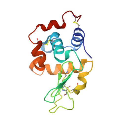
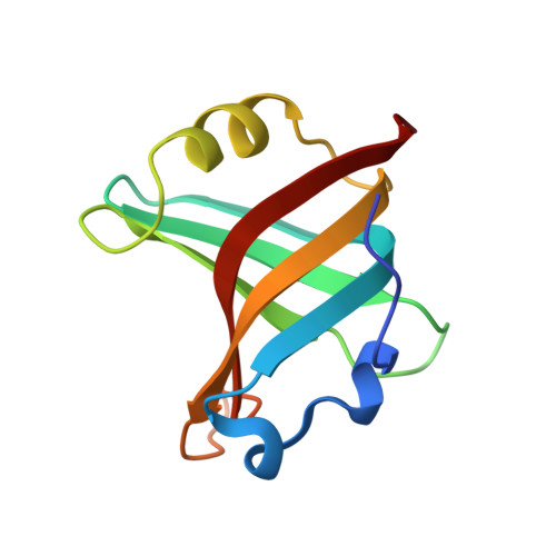
Tracking Molecular Recognition at the Atomic Level with a New Protein Scaffold Based on the OB-Fold.

Steemson, J.D.Baake, M.Rakonjac, J.Arcus, V.L.Liddament, M.T.

(2014) PLoS One 9: e86050-e86050

  • DOI: https://doi.org/10.1371/journal.pone.0086050
  • Primary Citation Related Structures: 
    4GLA, 4GLV, 4GN3, 4GN4, 4GN5

  • PubMed Abstract: 

    The OB-fold is a small, versatile single-domain protein binding module that occurs in all forms of life, where it binds protein, carbohydrate, nucleic acid and small-molecule ligands. We have exploited this natural plasticity to engineer a new class of non-immunoglobulin alternatives to antibodies with unique structural and biophysical characteristics. We present here the engineering of the OB-fold anticodon recognition domain from aspartyl tRNA synthetase taken from the thermophile Pyrobaculum aerophilum. For this single-domain scaffold we have coined the term OBody. Starting from a naïve combinatorial library, we engineered an OBody with 3 nM affinity for hen egg-white lysozyme, by optimising the affinity of a naïve OBody 11,700-fold over several affinity maturation steps, using phage display. At each maturation step a crystal structure of the engineered OBody in complex with hen egg-white lysozyme was determined, showing binding elements in atomic detail. These structures have given us an unprecedented insight into the directed evolution of affinity for a single antigen on the molecular scale. The engineered OBodies retain the high thermal stability of the parental OB-fold despite mutation of up to 22% of their residues. They can be expressed in soluble form and also purified from bacteria at high yields. They also lack disulfide bonds. These data demonstrate the potential of OBodies as a new scaffold for the engineering of specific binding reagents and provide a platform for further development of future OBody-based applications.


  • Organizational Affiliation
    • Department of Biological Sciences, University of Waikato, Hamilton, New Zealand.

Macromolecule Content 

  • Total Structure Weight: 107.32 kDa 
  • Atom Count: 7,893 
  • Modeled Residue Count: 941 
  • Deposited Residue Count: 944 
  • Unique protein chains: 2

Macromolecules

Find similar proteins by:|  3D Structure
Entity ID: 1
MoleculeChains  Sequence LengthOrganismDetailsImage
Lysozyme C
A, C, E, G
129Gallus gallusMutation(s): 0 
EC: 3.2.1.17
UniProt
Find proteins for P00698 (Gallus gallus)
Explore P00698 
Go to UniProtKB:  P00698
Entity Groups
Sequence Clusters30% Identity50% Identity70% Identity90% Identity95% Identity100% Identity
UniProt GroupP00698
Sequence Annotations
Expand
Reference Sequence
Find similar proteins by:|  3D Structure
Entity ID: 2
MoleculeChains  Sequence LengthOrganismDetailsImage
OBody AM3L09
B, D, F, H
107Pyrobaculum aerophilumMutation(s): 0 
Gene Names: aspS

Small Molecules

Ligands 4 Unique
IDChains Name / Formula / InChI Key2D Diagram3D Interactions
EPE

Query on EPE


Download:Ideal Coordinates CCD File
S [auth C]4-(2-HYDROXYETHYL)-1-PIPERAZINE ETHANESULFONIC ACID
C8 H18 N2 O4 S
JKMHFZQWWAIEOD-UHFFFAOYSA-N
SO4

Query on SO4


Download:Ideal Coordinates CCD File
W [auth E]SULFATE ION
O4 S
QAOWNCQODCNURD-UHFFFAOYSA-L
PO4

Query on PO4


Download:Ideal Coordinates CCD File
DA [auth G],
FA [auth H],
I [auth A],
J [auth A],
X [auth F]
PHOSPHATE ION
O4 P
NBIIXXVUZAFLBC-UHFFFAOYSA-K
GOL

Query on GOL


Download:Ideal Coordinates CCD File
AA [auth F]
BA [auth F]
CA [auth F]
EA [auth G]
GA [auth H]
AA [auth F],
BA [auth F],
CA [auth F],
EA [auth G],
GA [auth H],
HA [auth H],
IA [auth H],
K [auth A],
L [auth B],
M [auth B],
N [auth C],
O [auth C],
P [auth C],
Q [auth C],
R [auth C],
T [auth D],
U [auth E],
V [auth E],
Y [auth F],
Z [auth F]
GLYCEROL
C3 H8 O3
PEDCQBHIVMGVHV-UHFFFAOYSA-N

Experimental Data & Validation

Experimental Data

  • Method: X-RAY DIFFRACTION
  • Resolution: 2.57 Å
  • R-Value Free:  0.245 (Depositor), 0.235 (DCC) 
  • R-Value Work:  0.201 (Depositor), 0.191 (DCC) 
  • R-Value Observed: 0.204 (Depositor) 
Space Group: P 1
Unit Cell:
Length ( Å )Angle ( ˚ )
a = 59α = 72.17
b = 69.38β = 69.46
c = 76.36γ = 77.55
Software Package:
Software NamePurpose
MOSFLMdata reduction
SCALAdata scaling
PHASERphasing
PHENIXrefinement
PDB_EXTRACTdata extraction

Structure Validation

View Full Validation Report


Entry History 

Deposition Data

Revision History  (Full details and data files)

  • Version 1.0: 2013-08-21
    Type: Initial release
  • Version 1.1: 2013-08-28
    Changes: Experimental preparation
  • Version 1.2: 2014-02-12
    Changes: Database references
  • Version 1.3: 2024-10-16
    Changes: Data collection, Database references, Derived calculations, Structure summary