4HFC | pdb_00004hfc

The GLIC pentameric Ligand-Gated Ion Channel F14'A ethanol-sensitive mutant complexed to 2-bromo-ethanol


Experimental Data Snapshot

  • Method: X-RAY DIFFRACTION
  • Resolution: 3.05 Å
  • R-Value Free: 
    0.220 (Depositor), 0.218 (DCC) 
  • R-Value Work: 
    0.192 (Depositor) 
  • R-Value Observed: 
    0.193 (Depositor) 

wwPDB Validation 3D Report Full Report

Validation slider image for 4HFC

Ligand Structure Quality Assessment 


This is version 1.2 of the entry. See complete history

Literature

Structural basis for potentiation by alcohols and anaesthetics in a ligand-gated ion channel.

Sauguet, L.Howard, R.J.Malherbe, L.Lee, U.S.Corringer, P.J.Harris, R.A.Delarue, M.

(2013) Nat Commun 4: 1697-1697

  • DOI: https://doi.org/10.1038/ncomms2682
  • Primary Citation Related Structures: 
    4HFB, 4HFC, 4HFD, 4HFE, 4HFH

  • PubMed Abstract: 

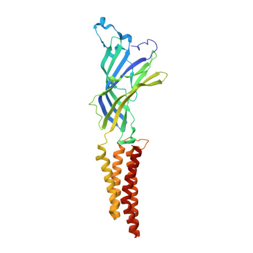
    Ethanol alters nerve signalling by interacting with proteins in the central nervous system, particularly pentameric ligand-gated ion channels. A recent series of mutagenesis experiments on Gloeobacter violaceus ligand-gated ion channel, a prokaryotic member of this family, identified a single-site variant that is potentiated by pharmacologically relevant concentrations of ethanol. Here we determine crystal structures of the ethanol-sensitized variant in the absence and presence of ethanol and related modulators, which bind in a transmembrane cavity between channel subunits and may stabilize the open form of the channel. Structural and mutagenesis studies defined overlapping mechanisms of potentiation by alcohols and anaesthetics via the inter-subunit cavity. Furthermore, homology modelling show this cavity to be conserved in human ethanol-sensitive glycine and GABA(A) receptors, and to involve residues previously shown to influence alcohol and anaesthetic action on these proteins. These results suggest a common structural basis for ethanol potentiation of an important class of targets for neurological actions of ethanol.


  • Organizational Affiliation
    • Unité de Dynamique Structurale des Macromolécules, Institut Pasteur, F-75015 Paris, France.

Macromolecule Content 

  • Total Structure Weight: 188.56 kDa 
  • Atom Count: 12,984 
  • Modeled Residue Count: 1,555 
  • Deposited Residue Count: 1,585 
  • Unique protein chains: 1

Macromolecules

Find similar proteins by:|  3D Structure
Entity ID: 1
MoleculeChains  Sequence LengthOrganismDetailsImage
Proton-gated ion channel
A, B, C, D, E
317Gloeobacter violaceus PCC 7421Mutation(s): 1 
Gene Names: glvIglr4197
Membrane Entity: Yes 
UniProt
Find proteins for Q7NDN8 (Gloeobacter violaceus (strain ATCC 29082 / PCC 7421))
Explore Q7NDN8 
Go to UniProtKB:  Q7NDN8
Entity Groups
Sequence Clusters30% Identity50% Identity70% Identity90% Identity95% Identity100% Identity
UniProt GroupQ7NDN8
Sequence Annotations
Expand
Reference Sequence

Small Molecules

Ligands 6 Unique
IDChains Name / Formula / InChI Key2D Diagram3D Interactions
PLC

Query on PLC


Download:Ideal Coordinates CCD File
FA [auth D],
M [auth A],
MA [auth E],
T [auth B],
Z [auth C]
DIUNDECYL PHOSPHATIDYL CHOLINE
C32 H65 N O8 P
IJFVSSZAOYLHEE-SSEXGKCCSA-O
LMT

Query on LMT


Download:Ideal Coordinates CCD File
AA [auth D]
F [auth A]
GA [auth E]
N [auth B]
U [auth C]
AA [auth D],
F [auth A],
GA [auth E],
N [auth B],
U [auth C],
V [auth C]
DODECYL-BETA-D-MALTOSIDE
C24 H46 O11
NLEBIOOXCVAHBD-QKMCSOCLSA-N
BRJ

Query on BRJ


Download:Ideal Coordinates CCD File
DA [auth D],
G [auth A],
KA [auth E],
P [auth B],
X [auth C]
2-BROMOETHANOL
C2 H5 Br O
LDLCZOVUSADOIV-UHFFFAOYSA-N
ACT

Query on ACT


Download:Ideal Coordinates CCD File
CA [auth D],
IA [auth E],
JA [auth E],
Q [auth B],
R [auth B]
ACETATE ION
C2 H3 O2
QTBSBXVTEAMEQO-UHFFFAOYSA-M
CL

Query on CL


Download:Ideal Coordinates CCD File
BA [auth D]
H [auth A]
HA [auth E]
I [auth A]
J [auth A]
BA [auth D],
H [auth A],
HA [auth E],
I [auth A],
J [auth A],
O [auth B],
W [auth C]
CHLORIDE ION
Cl
VEXZGXHMUGYJMC-UHFFFAOYSA-M
NA

Query on NA


Download:Ideal Coordinates CCD File
EA [auth D]
K [auth A]
L [auth A]
LA [auth E]
S [auth B]
EA [auth D],
K [auth A],
L [auth A],
LA [auth E],
S [auth B],
Y [auth C]
SODIUM ION
Na
FKNQFGJONOIPTF-UHFFFAOYSA-N

Experimental Data & Validation

Experimental Data

  • Method: X-RAY DIFFRACTION
  • Resolution: 3.05 Å
  • R-Value Free:  0.220 (Depositor), 0.218 (DCC) 
  • R-Value Work:  0.192 (Depositor) 
  • R-Value Observed: 0.193 (Depositor) 
Space Group: C 1 2 1
Unit Cell:
Length ( Å )Angle ( ˚ )
a = 180.88α = 90
b = 133.14β = 102.22
c = 159.35γ = 90
Software Package:
Software NamePurpose
BUSTERrefinement
XDSdata reduction
SCALAdata scaling

Structure Validation

View Full Validation Report


Ligand Structure Quality Assessment 


Entry History 

Deposition Data

Revision History  (Full details and data files)

  • Version 1.0: 2013-04-17
    Type: Initial release
  • Version 1.1: 2013-07-03
    Changes: Database references
  • Version 1.2: 2024-02-28
    Changes: Data collection, Database references, Derived calculations