4HZN | pdb_00004hzn

The Structure of the Bifunctional Acetyltransferase/Decarboxylase LnmK from the Leinamycin Biosynthetic Pathway Revealing Novel Activity for a Double Hot Dog Fold


Experimental Data Snapshot

  • Method: X-RAY DIFFRACTION
  • Resolution: 2.25 Å
  • R-Value Free: 
    0.254 (Depositor), 0.252 (DCC) 
  • R-Value Work: 
    0.200 (Depositor), 0.199 (DCC) 
  • R-Value Observed: 
    0.202 (Depositor) 

wwPDB Validation 3D Report Full Report

Validation slider image for 4HZN

This is version 1.2 of the entry. See complete history

Literature

Structure of the Bifunctional Acyltransferase/Decarboxylase LnmK from the Leinamycin Biosynthetic Pathway Revealing Novel Activity for a Double-Hot-Dog Fold.

Lohman, J.R.Bingman, C.A.Phillips, G.N.Shen, B.

(2013) Biochemistry 52: 902-911

  • DOI: https://doi.org/10.1021/bi301652y
  • Primary Citation Related Structures: 
    4HZN, 4HZO, 4HZP

  • PubMed Abstract: 

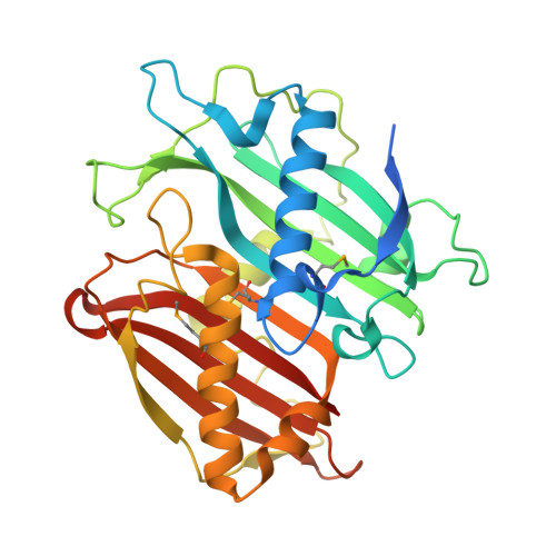
    The β-branched C3 unit in leinamycin biosynthesis is installed by a set of four proteins, LnmFKLM. In vitro biochemical investigation confirmed that LnmK is a bifunctional acyltransferase/decarboxylase (AT/DC) that catalyzes first self-acylation using methylmalonyl-CoA as a substrate and subsequently transacylation of the methylmalonyl group to the phosphopantetheinyl group of the LnmL acyl carrier protein [Liu, T., Huang, Y., and Shen, B. (2009) J. Am. Chem. Soc. 131, 6900-6901]. LnmK shows no sequence homology to proteins of known function, representing a new family of AT/DC enzymes. Here we report the X-ray structure of LnmK. LnmK is homodimer with each of the monomers adopting a double-hot-dog fold. Cocrystallization of LnmK with methylmalonyl-CoA revealed an active site tunnel terminated by residues from the dimer interface. In contrast to canonical AT and ketosynthase enzymes that employ Ser or Cys as an active site residue, none of these residues are found in the vicinity of the LnmK active site. Instead, three tyrosines were identified, one of which, Tyr62, was established, by site-directed mutagenesis, to be the most likely active site residue for the AT activity of LnmK. LnmK represents the first AT enzyme that employs a Tyr as an active site residue and the first member of the family of double-hot-dog fold enzymes that displays an AT activity known to date. The LnmK structure sets the stage for probing of the DC activity of LnmK through site-directed mutagenesis. These findings highlight natural product biosynthetic machinery as a rich source of novel enzyme activities, mechanisms, and structures.


  • Organizational Affiliation
    • Department of Chemistry, Scripps Research Institute, The Scripps Research Institute, Jupiter, Florida 33485, United States.

Macromolecule Content 

  • Total Structure Weight: 38.07 kDa 
  • Atom Count: 2,511 
  • Modeled Residue Count: 299 
  • Deposited Residue Count: 333 
  • Unique protein chains: 1

Macromolecules

Find similar proteins by:|  3D Structure
Entity ID: 1
MoleculeChains  Sequence LengthOrganismDetailsImage
Bifunctional Methylmalonyl-CoA:ACP Acyltransferase/Decarboxylase333Streptomyces atroolivaceusMutation(s): 0 
Gene Names: LnmK
UniProt
Find proteins for Q8GGP1 (Streptomyces atroolivaceus)
Explore Q8GGP1 
Go to UniProtKB:  Q8GGP1
Entity Groups
Sequence Clusters30% Identity50% Identity70% Identity90% Identity95% Identity100% Identity
UniProt GroupQ8GGP1
Sequence Annotations
Expand
Reference Sequence

Small Molecules

Ligands 3 Unique
IDChains Name / Formula / InChI Key2D Diagram3D Interactions
TRS

Query on TRS


Download:Ideal Coordinates CCD File
B [auth A]2-AMINO-2-HYDROXYMETHYL-PROPANE-1,3-DIOL
C4 H12 N O3
LENZDBCJOHFCAS-UHFFFAOYSA-O
SO4

Query on SO4


Download:Ideal Coordinates CCD File
D [auth A],
E [auth A],
F [auth A],
G [auth A],
H [auth A]
SULFATE ION
O4 S
QAOWNCQODCNURD-UHFFFAOYSA-L
GOL

Query on GOL


Download:Ideal Coordinates CCD File
C [auth A]GLYCEROL
C3 H8 O3
PEDCQBHIVMGVHV-UHFFFAOYSA-N
Modified Residues  1 Unique
IDChains TypeFormula2D DiagramParent
MSE
Query on MSE
A
L-PEPTIDE LINKINGC5 H11 N O2 SeMET

Experimental Data & Validation

Experimental Data

  • Method: X-RAY DIFFRACTION
  • Resolution: 2.25 Å
  • R-Value Free:  0.254 (Depositor), 0.252 (DCC) 
  • R-Value Work:  0.200 (Depositor), 0.199 (DCC) 
  • R-Value Observed: 0.202 (Depositor) 
Space Group: P 61 2 2
Unit Cell:
Length ( Å )Angle ( ˚ )
a = 60.165α = 90
b = 60.165β = 90
c = 311.193γ = 120
Software Package:
Software NamePurpose
HKL-2000data collection
SOLVEphasing
REFMACrefinement
HKL-2000data reduction
SCALEPACKdata scaling

Structure Validation

View Full Validation Report


Entry History 

Deposition Data

Revision History  (Full details and data files)

  • Version 1.0: 2013-01-30
    Type: Initial release
  • Version 1.1: 2013-02-20
    Changes: Database references
  • Version 1.2: 2024-11-27
    Changes: Data collection, Database references, Derived calculations, Structure summary