4J9A

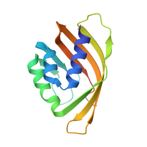
Engineered Digoxigenin binder DIG10.3


Experimental Data Snapshot

  • Method: X-RAY DIFFRACTION
  • Resolution: 3.20 Å
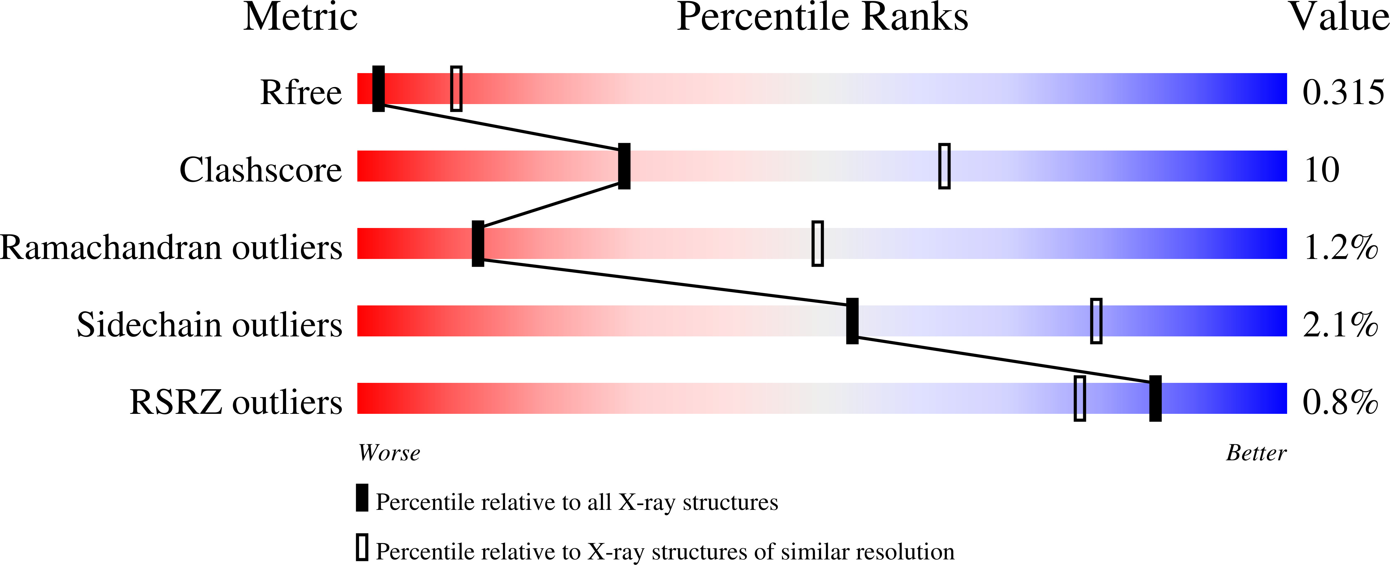
  • R-Value Free: 0.317 
  • R-Value Work: 0.262 
  • R-Value Observed: 0.265 

wwPDB Validation   3D Report Full Report


Ligand Structure Quality Assessment 


This is version 1.5 of the entry. See complete history


Literature

Computational design of ligand-binding proteins with high affinity and selectivity.

Tinberg, C.E.Khare, S.D.Dou, J.Doyle, L.Nelson, J.W.Schena, A.Jankowski, W.Kalodimos, C.G.Johnsson, K.Stoddard, B.L.Baker, D.

(2013) Nature 501: 212-216

  • DOI: https://doi.org/10.1038/nature12443
  • Primary Citation of Related Structures:  
    4J8T, 4J9A

  • PubMed Abstract: 

    The ability to design proteins with high affinity and selectivity for any given small molecule is a rigorous test of our understanding of the physiochemical principles that govern molecular recognition. Attempts to rationally design ligand-binding proteins have met with little success, however, and the computational design of protein-small-molecule interfaces remains an unsolved problem. Current approaches for designing ligand-binding proteins for medical and biotechnological uses rely on raising antibodies against a target antigen in immunized animals and/or performing laboratory-directed evolution of proteins with an existing low affinity for the desired ligand, neither of which allows complete control over the interactions involved in binding. Here we describe a general computational method for designing pre-organized and shape complementary small-molecule-binding sites, and use it to generate protein binders to the steroid digoxigenin (DIG). Of seventeen experimentally characterized designs, two bind DIG; the model of the higher affinity binder has the most energetically favourable and pre-organized interface in the design set. A comprehensive binding-fitness landscape of this design, generated by library selections and deep sequencing, was used to optimize its binding affinity to a picomolar level, and X-ray co-crystal structures of two variants show atomic-level agreement with the corresponding computational models. The optimized binder is selective for DIG over the related steroids digitoxigenin, progesterone and β-oestradiol, and this steroid binding preference can be reprogrammed by manipulation of explicitly designed hydrogen-bonding interactions. The computational design method presented here should enable the development of a new generation of biosensors, therapeutics and diagnostics.


  • Organizational Affiliation

    Department of Biochemistry, University of Washington, Seattle, WA 98195, USA.


Macromolecules
Find similar proteins by:  (by identity cutoff)  |  3D Structure
Entity ID: 1
MoleculeChains Sequence LengthOrganismDetailsImage
Engineered Digoxigenin binder protein DIG10.3
A, B, C, D, E
A, B, C, D, E, F, G, H, I
137Pseudomonas aeruginosa PAO1Mutation(s): 20 
Gene Names: PA3332
UniProt
Find proteins for Q9HYR3 (Pseudomonas aeruginosa (strain ATCC 15692 / DSM 22644 / CIP 104116 / JCM 14847 / LMG 12228 / 1C / PRS 101 / PAO1))
Explore Q9HYR3 
Go to UniProtKB:  Q9HYR3
Entity Groups  
Sequence Clusters30% Identity50% Identity70% Identity90% Identity95% Identity100% Identity
UniProt GroupQ9HYR3
Sequence Annotations
Expand
  • Reference Sequence
Small Molecules
Ligands 1 Unique
IDChains Name / Formula / InChI Key2D Diagram3D Interactions
DOG
Query on DOG

Download Ideal Coordinates CCD File 
J [auth A]
K [auth B]
L [auth C]
M [auth D]
N [auth E]
J [auth A],
K [auth B],
L [auth C],
M [auth D],
N [auth E],
O [auth F],
P [auth G],
Q [auth H],
R [auth I]
DIGOXIGENIN
C23 H34 O5
SHIBSTMRCDJXLN-KCZCNTNESA-N
Binding Affinity Annotations 
IDSourceBinding Affinity
DOG PDBBind:  4J9A Kd: 6 (nM) from 1 assay(s)
BindingDB:  4J9A Kd: 474 (nM) from 1 assay(s)
Experimental Data & Validation

Experimental Data

  • Method: X-RAY DIFFRACTION
  • Resolution: 3.20 Å
  • R-Value Free: 0.317 
  • R-Value Work: 0.262 
  • R-Value Observed: 0.265 
  • Space Group: C 1 2 1
Unit Cell:
Length ( Å )Angle ( ˚ )
a = 132.793α = 90
b = 90.993β = 92.68
c = 110.075γ = 90
Software Package:
Software NamePurpose
DENZOdata reduction
SCALEPACKdata scaling
REFMACrefinement
PDB_EXTRACTdata extraction
BOSdata collection
HKL-2000data reduction
HKL-2000data scaling
PHASERphasing

Structure Validation

View Full Validation Report


Ligand Structure Quality Assessment 


Entry History 

Deposition Data

Revision History  (Full details and data files)

  • Version 1.0: 2013-06-26
    Type: Initial release
  • Version 1.1: 2013-08-14
    Changes: Database references
  • Version 1.2: 2013-09-18
    Changes: Database references
  • Version 1.3: 2013-09-25
    Changes: Database references
  • Version 1.4: 2017-11-15
    Changes: Refinement description
  • Version 1.5: 2024-02-28
    Changes: Data collection, Database references, Derived calculations, Refinement description