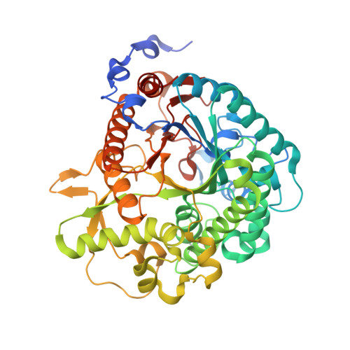
4JIE

Structural analysis and insights into glycon specificity of the rice GH1 Os7BGlu26 beta-D-mannosidase


Experimental Data Snapshot

  • Method: X-RAY DIFFRACTION
  • Resolution: 2.45 Å
  • R-Value Free: 0.195 
  • R-Value Work: 0.153 
  • R-Value Observed: 0.155 

Starting Model: experimental
View more details

wwPDB Validation   3D Report Full Report


Ligand Structure Quality Assessment 


This is version 1.4 of the entry. See complete history


Literature

Structural analysis and insights into the glycon specificity of the rice GH1 Os7BGlu26 beta-D-mannosidase

Tankrathok, A.Iglesias-Fernandez, J.Luang, S.Robinson, R.C.Kimura, A.Rovira, C.Hrmova, M.Ketudat Cairns, J.R.

(2013) Acta Crystallogr D Biol Crystallogr 69: 2124-2135

  • DOI: https://doi.org/10.1107/S0907444913020568
  • Primary Citation of Related Structures:  
    4JHO, 4JIE

  • PubMed Abstract: 

    Rice Os7BGlu26 is a GH1 family glycoside hydrolase with a threefold higher kcat/Km value for 4-nitrophenyl β-D-mannoside (4NPMan) compared with 4-nitrophenyl β-D-glucoside (4NPGlc). To investigate its selectivity for β-D-mannoside and β-D-glucoside substrates, the structures of apo Os7BGlu26 at a resolution of 2.20 Å and of Os7BGlu26 with mannose at a resolution of 2.45 Å were elucidated from isomorphous crystals in space group P212121. The (β/α)8-barrel structure is similar to other GH1 family structures, but with a narrower active-site cleft. The Os7BGlu26 structure with D-mannose corresponds to a product complex, with β-D-mannose in the (1)S5 skew-boat conformation. Docking of the (1)S3, (1)S5, (2)SO and (3)S1 pyranose-ring conformations of 4NPMan and 4NPGlc substrates into the active site of Os7BGlu26 indicated that the lowest energies were in the (1)S5 and (1)S3 skew-boat conformations. Comparison of these docked conformers with other rice GH1 structures revealed differences in the residues interacting with the catalytic acid/base between enzymes with and without β-D-mannosidase activity. The mutation of Tyr134 to Trp in Os7BGlu26 resulted in similar kcat/Km values for 4NPMan and 4NPGlc, while mutation of Tyr134 to Phe resulted in a 37-fold higher kcat/Km for 4NPMan than 4NPGlc. Mutation of Cys182 to Thr decreased both the activity and the selectivity for β-D-mannoside. It was concluded that interactions with the catalytic acid/base play a significant role in glycon selection.


  • Organizational Affiliation

    School of Biochemistry, Institute of Science, Suranaree University of Technology, Nakhon Ratchasima 30000, Thailand.


Macromolecules
Find similar proteins by:  (by identity cutoff)  |  3D Structure
Entity ID: 1
MoleculeChains Sequence LengthOrganismDetailsImage
Beta-mannosidase/beta-glucosidase503Oryza sativa Indica GroupMutation(s): 0 
EC: 3.2.1.25
UniProt
Find proteins for A2YPH1 (Oryza sativa subsp. indica)
Explore A2YPH1 
Go to UniProtKB:  A2YPH1
Entity Groups  
Sequence Clusters30% Identity50% Identity70% Identity90% Identity95% Identity100% Identity
UniProt GroupA2YPH1
Sequence Annotations
Expand
  • Reference Sequence
Small Molecules
Ligands 3 Unique
IDChains Name / Formula / InChI Key2D Diagram3D Interactions
EPE
Query on EPE

Download Ideal Coordinates CCD File 
H [auth A]4-(2-HYDROXYETHYL)-1-PIPERAZINE ETHANESULFONIC ACID
C8 H18 N2 O4 S
JKMHFZQWWAIEOD-UHFFFAOYSA-N
BMA
Query on BMA

Download Ideal Coordinates CCD File 
I [auth A]beta-D-mannopyranose
C6 H12 O6
WQZGKKKJIJFFOK-RWOPYEJCSA-N
GOL
Query on GOL

Download Ideal Coordinates CCD File 
B [auth A]
C [auth A]
D [auth A]
E [auth A]
F [auth A]
B [auth A],
C [auth A],
D [auth A],
E [auth A],
F [auth A],
G [auth A]
GLYCEROL
C3 H8 O3
PEDCQBHIVMGVHV-UHFFFAOYSA-N
Experimental Data & Validation

Experimental Data

  • Method: X-RAY DIFFRACTION
  • Resolution: 2.45 Å
  • R-Value Free: 0.195 
  • R-Value Work: 0.153 
  • R-Value Observed: 0.155 
  • Space Group: P 21 21 21
Unit Cell:
Length ( Å )Angle ( ˚ )
a = 68.01α = 90
b = 73.624β = 90
c = 133.984γ = 90
Software Package:
Software NamePurpose
ADSCdata collection
MOLREPphasing
REFMACrefinement
HKL-2000data reduction
HKL-2000data scaling

Structure Validation

View Full Validation Report


Ligand Structure Quality Assessment 


Entry History 

Deposition Data

Revision History  (Full details and data files)

  • Version 1.0: 2013-10-09
    Type: Initial release
  • Version 1.1: 2014-04-23
    Changes: Database references
  • Version 1.2: 2020-07-29
    Type: Remediation
    Reason: Carbohydrate remediation
    Changes: Data collection, Database references, Derived calculations, Structure summary
  • Version 1.3: 2023-11-08
    Changes: Data collection, Database references, Refinement description, Structure summary
  • Version 1.4: 2024-11-20
    Changes: Structure summary