4K5O | pdb_00004k5o

Phosphonic Arginine Mimetics as Inhibitors of the M1 Aminopeptidases from Plasmodium falciparum


Experimental Data Snapshot

  • Method: X-RAY DIFFRACTION
  • Resolution: 1.90 Å
  • R-Value Free: 
    0.241 (Depositor), 0.239 (DCC) 
  • R-Value Work: 
    0.182 (Depositor), 0.181 (DCC) 
  • R-Value Observed: 
    0.185 (Depositor) 

Starting Model: experimental
View more details

wwPDB Validation 3D Report Full Report

Validation slider image for 4K5O

Ligand Structure Quality Assessment 


This is version 1.2 of the entry. See complete history

Literature

Synthesis and Structure-Activity Relationships of Phosphonic Arginine Mimetics as Inhibitors of the M1 and M17 Aminopeptidases from Plasmodium falciparum.

Kannan Sivaraman, K.Paiardini, A.Sienczyk, M.Ruggeri, C.Oellig, C.A.Dalton, J.P.Scammells, P.J.Drag, M.McGowan, S.

(2013) J Med Chem 56: 5213-5217

  • DOI: https://doi.org/10.1021/jm4005972
  • Primary Citation Related Structures: 
    4K3N, 4K5L, 4K5M, 4K5N, 4K5O, 4K5P

  • PubMed Abstract: 

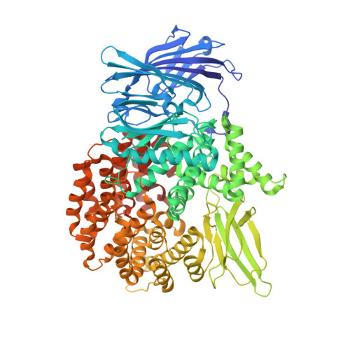
    The malaria parasite Plasmodium falciparum employs two metallo-aminopeptidases, PfA-M1 and PfA-M17, which are essential for parasite survival. Compounds that inhibit the activity of either enzyme represent leads for the development of new antimalarial drugs. Here we report the synthesis and structure-activity relationships of a small library of phosphonic acid arginine mimetics that probe the S1 pocket of both enzymes and map the necessary interactions that would be important for a dual inhibitor.


  • Organizational Affiliation
    • Department of Biochemistry and Molecular Biology, Monash University, Clayton Campus, Melbourne, VIC 3800, Australia.

Macromolecule Content 

  • Total Structure Weight: 105.01 kDa 
  • Atom Count: 8,279 
  • Modeled Residue Count: 889 
  • Deposited Residue Count: 895 
  • Unique protein chains: 1

Macromolecules

Find similar proteins by:|  3D Structure
Entity ID: 1
MoleculeChains  Sequence LengthOrganismDetailsImage
M1 family aminopeptidase895Plasmodium falciparum FcB1/ColumbiaMutation(s): 7 
EC: 3.4.11
UniProt
Find proteins for O96935 (Plasmodium falciparum (isolate 3D7))
Explore O96935 
Go to UniProtKB:  O96935
Entity Groups
Sequence Clusters30% Identity50% Identity70% Identity90% Identity95% Identity100% Identity
UniProt GroupO96935
Sequence Annotations
Expand
Reference Sequence

Small Molecules

Ligands 4 Unique
IDChains Name / Formula / InChI Key2D Diagram3D Interactions
1OT

Query on 1OT


Download:Ideal Coordinates CCD File
G [auth A]{(R)-amino[4-(1H-pyrazol-1-yl)phenyl]methyl}phosphonic acid
C10 H12 N3 O3 P
KEKSMECJAGVZSC-SNVBAGLBSA-N
GOL

Query on GOL


Download:Ideal Coordinates CCD File
B [auth A],
F [auth A]
GLYCEROL
C3 H8 O3
PEDCQBHIVMGVHV-UHFFFAOYSA-N
ZN

Query on ZN


Download:Ideal Coordinates CCD File
D [auth A]ZINC ION
Zn
PTFCDOFLOPIGGS-UHFFFAOYSA-N
MG

Query on MG


Download:Ideal Coordinates CCD File
C [auth A],
E [auth A]
MAGNESIUM ION
Mg
JLVVSXFLKOJNIY-UHFFFAOYSA-N

Experimental Data & Validation

Experimental Data

  • Method: X-RAY DIFFRACTION
  • Resolution: 1.90 Å
  • R-Value Free:  0.241 (Depositor), 0.239 (DCC) 
  • R-Value Work:  0.182 (Depositor), 0.181 (DCC) 
  • R-Value Observed: 0.185 (Depositor) 
Space Group: P 21 21 21
Unit Cell:
Length ( Å )Angle ( ˚ )
a = 76.056α = 90
b = 109.35β = 90
c = 118.518γ = 90
Software Package:
Software NamePurpose
REFMACrefinement
PDB_EXTRACTdata extraction
Blu-Icedata collection
XDSdata reduction
SCALAdata scaling
PHASERphasing

Structure Validation

View Full Validation Report


Ligand Structure Quality Assessment 


Entry History 

Deposition Data

  • Released Date: 2013-06-12 
  • Deposition Author(s): McGowan, S.

Revision History  (Full details and data files)

  • Version 1.0: 2013-06-12
    Type: Initial release
  • Version 1.1: 2013-07-24
    Changes: Database references
  • Version 1.2: 2023-09-20
    Changes: Data collection, Database references, Derived calculations, Refinement description