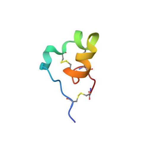
4LFQ

High resolution x-ray crystal structure of L-ShK toxin


Experimental Data Snapshot

  • Method: X-RAY DIFFRACTION
  • Resolution: 1.06 Å
  • R-Value Free: 0.160 
  • R-Value Work: 0.131 
  • R-Value Observed: 0.132 

wwPDB Validation   3D Report Full Report


This is version 1.4 of the entry. See complete history


Literature

Native Chemical Ligation at Asx-Cys, Glx-Cys: Chemical Synthesis and High-Resolution X-ray Structure of ShK Toxin by Racemic Protein Crystallography.

Dang, B.Kubota, T.Mandal, K.Bezanilla, F.Kent, S.B.

(2013) J Am Chem Soc 135: 11911-11919

  • DOI: https://doi.org/10.1021/ja4046795
  • Primary Citation of Related Structures:  
    4LFQ, 4LFS

  • PubMed Abstract: 

    We have re-examined the utility of native chemical ligation at -Gln/Glu-Cys- [Glx-Cys] and -Asn/Asp-Cys- [Asx-Cys] sites. Using the improved thioaryl catalyst 4-mercaptophenylacetic acid (MPAA), native chemical ligation could be performed at -Gln-Cys- and Asn-Cys- sites without side reactions. After optimization, ligation at a -Glu-Cys- site could also be used as a ligation site, with minimal levels of byproduct formation. However, -Asp-Cys- is not appropriate for use as a site for native chemical ligation because of formation of significant amounts of β-linked byproduct. The feasibility of native chemical ligation at -Gln-Cys- enabled a convergent total chemical synthesis of the enantiomeric forms of the ShK toxin protein molecule. The D-ShK protein molecule was ~50,000-fold less active in blocking the Kv1.3 channel than the L-ShK protein molecule. Racemic protein crystallography was used to obtain high-resolution X-ray diffraction data for ShK toxin. The structure was solved by direct methods and showed significant differences from the previously reported NMR structures in some regions of the ShK protein molecule.


  • Organizational Affiliation

    Department of Chemistry, Institute for Biophysical Dynamics, University of Chicago, Chicago, Illinois 60637, USA.


Macromolecules
Find similar proteins by:  (by identity cutoff)  |  3D Structure
Entity ID: 1
MoleculeChains Sequence LengthOrganismDetailsImage
Potassium channel toxin ShK35Stichodactyla helianthusMutation(s): 0 
UniProt
Find proteins for P29187 (Stichodactyla helianthus)
Explore P29187 
Go to UniProtKB:  P29187
Entity Groups  
Sequence Clusters30% Identity50% Identity70% Identity90% Identity95% Identity100% Identity
UniProt GroupP29187
Sequence Annotations
Expand
  • Reference Sequence
Experimental Data & Validation

Experimental Data

  • Method: X-RAY DIFFRACTION
  • Resolution: 1.06 Å
  • R-Value Free: 0.160 
  • R-Value Work: 0.131 
  • R-Value Observed: 0.132 
  • Space Group: C 2 2 21
Unit Cell:
Length ( Å )Angle ( ˚ )
a = 41.161α = 90
b = 49.491β = 90
c = 27.77γ = 90
Software Package:
Software NamePurpose
DENZOdata reduction
SCALEPACKdata scaling
PHASERphasing
PHENIXrefinement
PDB_EXTRACTdata extraction
HKL-2000data reduction
HKL-2000data scaling

Structure Validation

View Full Validation Report


Entry History 

Deposition Data

Revision History  (Full details and data files)

  • Version 1.0: 2013-08-14
    Type: Initial release
  • Version 1.1: 2013-09-04
    Changes: Database references
  • Version 1.2: 2017-11-15
    Changes: Refinement description
  • Version 1.3: 2023-08-02
    Changes: Database references, Derived calculations
  • Version 1.4: 2024-11-20
    Changes: Data collection, Structure summary