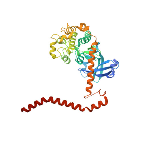
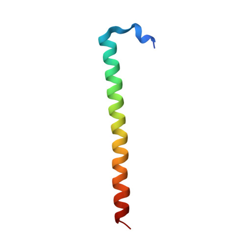
4LGD

Structural Basis for Autoactivation of Human Mst2 Kinase and Its Regulation by RASSF5


Experimental Data Snapshot

  • Method: X-RAY DIFFRACTION
  • Resolution: 3.05 Å
  • R-Value Free: 0.244 
  • R-Value Work: 0.199 
  • R-Value Observed: 0.202 

Starting Model: experimental
View more details

wwPDB Validation   3D Report Full Report


Ligand Structure Quality Assessment 


This is version 1.5 of the entry. See complete history


Literature

Structural Basis for Autoactivation of Human Mst2 Kinase and Its Regulation by RASSF5.

Ni, L.Li, S.Yu, J.Min, J.Brautigam, C.A.Tomchick, D.R.Pan, D.Luo, X.

(2013) Structure 21: 1757-1768

  • DOI: https://doi.org/10.1016/j.str.2013.07.008
  • Primary Citation of Related Structures:  
    4LG4, 4LGD

  • PubMed Abstract: 

    The tumor-suppressive Hippo pathway controls tissue homeostasis through balancing cell proliferation and apoptosis. Activation of the kinases Mst1 and Mst2 (Mst1/2) is a key upstream event in this pathway and remains poorly understood. Mst1/2 and their critical regulators RASSFs contain Salvador/RASSF1A/Hippo (SARAH) domains that can homo- and heterodimerize. Here, we report the crystal structures of human Mst2 alone and bound to RASSF5. Mst2 undergoes activation through transautophosphorylation at its activation loop, which requires SARAH-mediated homodimerization. RASSF5 disrupts Mst2 homodimer and blocks Mst2 autoactivation. Binding of RASSF5 to already activated Mst2, however, does not inhibit its kinase activity. Thus, RASSF5 can act as an inhibitor or a potential positive regulator of Mst2, depending on whether it binds to Mst2 before or after activation-loop phosphorylation. We propose that these temporally sensitive functions of RASSFs enable the Hippo pathway to respond to and integrate diverse cellular signals.


  • Organizational Affiliation

    Department of Pharmacology, The University of Texas Southwestern Medical Center, 6001 Forest Park Road, Dallas, TX 75390, USA.


Macromolecules
Find similar proteins by:  (by identity cutoff)  |  3D Structure
Entity ID: 1
MoleculeChains Sequence LengthOrganismDetailsImage
Serine/threonine-protein kinase 3
A, B, C, D
378Homo sapiensMutation(s): 1 
Gene Names: STK3KRS1MST2
EC: 2.7.11.1
UniProt & NIH Common Fund Data Resources
Find proteins for Q13188 (Homo sapiens)
Explore Q13188 
Go to UniProtKB:  Q13188
PHAROS:  Q13188
GTEx:  ENSG00000104375 
Entity Groups  
Sequence Clusters30% Identity50% Identity70% Identity90% Identity95% Identity100% Identity
UniProt GroupQ13188
Sequence Annotations
Expand
  • Reference Sequence
Find similar proteins by:  (by identity cutoff)  |  3D Structure
Entity ID: 2
MoleculeChains Sequence LengthOrganismDetailsImage
Ras association domain family member 5, RASSF5
E, F, G, H
49Homo sapiensMutation(s): 0 
Gene Names: RASSF5NORE1RAPL
UniProt & NIH Common Fund Data Resources
Find proteins for Q8WWW0 (Homo sapiens)
Explore Q8WWW0 
Go to UniProtKB:  Q8WWW0
PHAROS:  Q8WWW0
GTEx:  ENSG00000266094 
Entity Groups  
Sequence Clusters30% Identity50% Identity70% Identity90% Identity95% Identity100% Identity
UniProt GroupQ8WWW0
Sequence Annotations
Expand
  • Reference Sequence
Small Molecules
Ligands 4 Unique
IDChains Name / Formula / InChI Key2D Diagram3D Interactions
ANP
Query on ANP

Download Ideal Coordinates CCD File 
I [auth A],
L [auth B],
R [auth C],
W [auth D]
PHOSPHOAMINOPHOSPHONIC ACID-ADENYLATE ESTER
C10 H17 N6 O12 P3
PVKSNHVPLWYQGJ-KQYNXXCUSA-N
SO4
Query on SO4

Download Ideal Coordinates CCD File 
AA [auth D]
K [auth A]
N [auth B]
O [auth B]
P [auth B]
AA [auth D],
K [auth A],
N [auth B],
O [auth B],
P [auth B],
Q [auth B],
T [auth C],
Y [auth D],
Z [auth D]
SULFATE ION
O4 S
QAOWNCQODCNURD-UHFFFAOYSA-L
MG
Query on MG

Download Ideal Coordinates CCD File 
J [auth A],
M [auth B],
S [auth C],
X [auth D]
MAGNESIUM ION
Mg
JLVVSXFLKOJNIY-UHFFFAOYSA-N
NA
Query on NA

Download Ideal Coordinates CCD File 
BA [auth D],
U [auth C],
V [auth C]
SODIUM ION
Na
FKNQFGJONOIPTF-UHFFFAOYSA-N
Experimental Data & Validation

Experimental Data

  • Method: X-RAY DIFFRACTION
  • Resolution: 3.05 Å
  • R-Value Free: 0.244 
  • R-Value Work: 0.199 
  • R-Value Observed: 0.202 
  • Space Group: C 1 2 1
Unit Cell:
Length ( Å )Angle ( ˚ )
a = 123.403α = 90
b = 237.136β = 100.7
c = 95.862γ = 90
Software Package:
Software NamePurpose
DENZOdata reduction
SCALEPACKdata scaling
PHENIXrefinement
PDB_EXTRACTdata extraction
PHASESphasing

Structure Validation

View Full Validation Report


Ligand Structure Quality Assessment 


Entry History 

Deposition Data

Revision History  (Full details and data files)

  • Version 1.0: 2013-09-18
    Type: Initial release
  • Version 1.1: 2013-10-30
    Changes: Database references
  • Version 1.2: 2017-08-09
    Changes: Source and taxonomy
  • Version 1.3: 2017-11-15
    Changes: Refinement description
  • Version 1.4: 2018-02-28
    Changes: Data collection, Experimental preparation, Refinement description
  • Version 1.5: 2023-09-20
    Changes: Data collection, Database references, Derived calculations, Refinement description