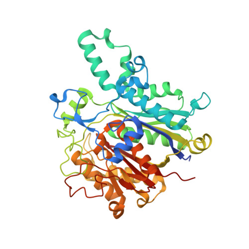
Structural insights into bacterial resistance to cerulenin.
Trajtenberg, F., Altabe, S., Larrieux, N., Ficarra, F., de Mendoza, D., Buschiazzo, A., Schujman, G.E.(2014) FEBS J 281: 2324-2338
- PubMed: 24641521
- DOI: https://doi.org/10.1111/febs.12785
- Primary Citation of Related Structures:
4LS5, 4LS6, 4LS7, 4LS8 - PubMed Abstract:
Cerulenin is a fungal toxin that inhibits both eukaryotic and prokaryotic ketoacyl-acyl carrier protein synthases or condensing enzymes. It has been used experimentally to treat cancer and obesity, and is a potent inhibitor of bacterial growth. Understanding the molecular mechanisms of resistance to cerulenin and similar compounds is thus highly relevant for human health. We have previously described a Bacillus subtilis cerulenin-resistant strain, expressing a point-mutated condensing enzyme FabF (FabF[I108F]) (i.e. FabF with isoleucine 108 substituted by phenylalanine). We now report the crystal structures of wild-type FabF from B. subtilis, both alone and in complex with cerulenin, as well as of the FabF[I108F] mutant protein. The three-dimensional structure of FabF[I108F] constitutes the first atomic model of a condensing enzyme that remains active in the presence of the inhibitor. Soaking the mycotoxin into preformed wild-type FabF crystals allowed for noncovalent binding into its specific pocket within the FabF core. Interestingly, only co-crystallization experiments allowed us to trap the covalent complex. Our structure shows that the covalent bond between Cys163 and cerulenin, in contrast to that previously proposed, implicates carbon C3 of the inhibitor. The similarities between Escherichia coli and B. subtilis FabF structures did not explain the reported inability of ecFabF[I108F] (i.e. FabF from Escherichia coli with isoleucine 108 substituted by phenylalanine) to elongate medium and long-chain acyl-ACPs. We now demonstrate that the E. coli modified enzyme efficiently catalyzes the synthesis of medium and long-chain ketoacyl-ACPs. We also characterized another cerulenin-insensitive form of FabF, conferring a different phenotype in B. subtilis. The structural, biochemical and physiological data presented, shed light on the mechanisms of FabF catalysis and resistance to cerulenin. Crystallographic data (including atomic coordinates and structure factors) have been deposited in the Protein Data Bank under accession codes 4LS5, 4LS6, 4LS7 and 4LS8.
- Institut Pasteur de Montevideo, Unit of Protein Crystallography, Montevideo, Uruguay.
Organizational Affiliation:


