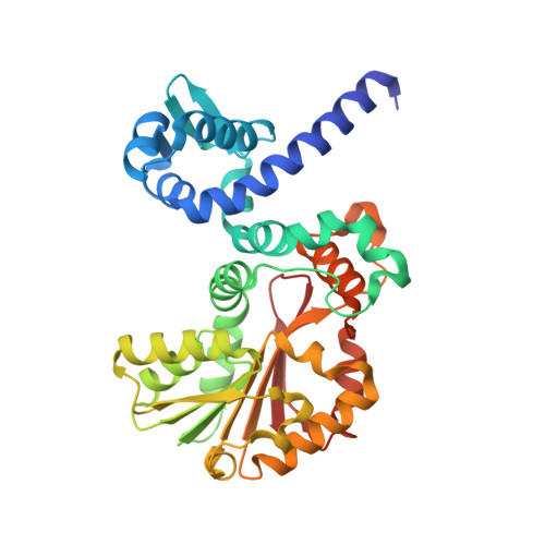
4M71

Mutant structure of methyltransferase from Streptomyces hygroscopicus complexed with S-adenosyl-L-homocysteine and methylphenylpyruvic acid


Experimental Data Snapshot

  • Method: X-RAY DIFFRACTION
  • Resolution: 2.21 Å
  • R-Value Free: 0.229 
  • R-Value Work: 0.158 
  • R-Value Observed: 0.162 

Starting Model: experimental
View more details

wwPDB Validation   3D Report Full Report


Ligand Structure Quality Assessment 


This is version 2.0 of the entry. See complete history


Literature

Structure and mechanism of a nonhaem-iron SAM-dependent C-methyltransferase and its engineering to a hydratase and an O-methyltransferase

Zou, X.W.Liu, Y.C.Hsu, N.S.Huang, C.J.Lyu, S.Y.Chan, H.C.Chang, C.Y.Yeh, H.W.Lin, K.H.Wu, C.J.Tsai, M.D.Li, T.L.

(2014) Acta Crystallogr D Biol Crystallogr 70: 1549-1560

  • DOI: https://doi.org/10.1107/S1399004714005239
  • Primary Citation of Related Structures:  
    4KIB, 4KIC, 4KIF, 4KIG, 4M6X, 4M6Y, 4M71, 4M72, 4M73, 4M74

  • PubMed Abstract: 

    In biological systems, methylation is most commonly performed by methyltransferases (MTs) using the electrophilic methyl source S-adenosyl-L-methionine (SAM) via the S(N)2 mechanism. (2S,3S)-β-Methylphenylalanine, a nonproteinogenic amino acid, is a building unit of the glycopeptide antibiotic mannopeptimycin. The gene product of mppJ from the mannopeptimycin-biosynthetic gene cluster is the MT that methylates the benzylic C atom of phenylpyruvate (Ppy) to give βMePpy. Although the benzylic C atom of Ppy is acidic, how its nucleophilicity is further enhanced to become an acceptor for C-methylation has not conclusively been determined. Here, a structural approach is used to address the mechanism of MppJ and to engineer it for new functions. The purified MppJ displays a turquoise colour, implying the presence of a metal ion. The crystal structures reveal MppJ to be the first ferric ion SAM-dependent MT. An additional four structures of binary and ternary complexes illustrate the molecular mechanism for the metal ion-dependent methyltransfer reaction. Overall, MppJ has a nonhaem iron centre that bind, orients and activates the α-ketoacid substrate and has developed a sandwiched bi-water device to avoid the formation of the unwanted reactive oxo-iron(IV) species during the C-methylation reaction. This discovery further prompted the conversion of the MT into a structurally/functionally unrelated new enzyme. Through stepwise mutagenesis and manipulation of coordination chemistry, MppJ was engineered to perform both Lewis acid-assisted hydration and/or O-methyltransfer reactions to give stereospecific new compounds. This process was validated by six crystal structures. The results reported in this study will facilitate the development and design of new biocatalysts for difficult-to-synthesize biochemicals.


  • Organizational Affiliation

    Genomics Research Center, Academia Sinica, Taipei 115, Taiwan.


Macromolecules
Find similar proteins by:  (by identity cutoff)  |  3D Structure
Entity ID: 1
MoleculeChains Sequence LengthOrganismDetailsImage
Methyltransferase MppJ
A, B
337Streptomyces hygroscopicusMutation(s): 2 
Gene Names: mppJ
EC: 2.1.1 (PDB Primary Data), 2.1.1.281 (UniProt)
UniProt
Find proteins for Q643C8 (Streptomyces hygroscopicus)
Explore Q643C8 
Go to UniProtKB:  Q643C8
Entity Groups  
Sequence Clusters30% Identity50% Identity70% Identity90% Identity95% Identity100% Identity
UniProt GroupQ643C8
Sequence Annotations
Expand
  • Reference Sequence
Small Molecules
Ligands 6 Unique
IDChains Name / Formula / InChI Key2D Diagram3D Interactions
SAH
Query on SAH

Download Ideal Coordinates CCD File 
C [auth A],
Q [auth B]
S-ADENOSYL-L-HOMOCYSTEINE
C14 H20 N6 O5 S
ZJUKTBDSGOFHSH-WFMPWKQPSA-N
56D
Query on 56D

Download Ideal Coordinates CCD File 
D [auth A],
R [auth B]
(3R)-2-oxo-3-phenylbutanoic acid
C10 H10 O3
AXLLOSUYAVXOIN-SSDOTTSWSA-N
HF2
Query on HF2

Download Ideal Coordinates CCD File 
F [auth A],
T [auth B]
(2R)-2-hydroxy-3-phenylpropanoic acid
C9 H10 O3
VOXXWSYKYCBWHO-MRVPVSSYSA-N
IOD
Query on IOD

Download Ideal Coordinates CCD File 
P [auth A]IODIDE ION
I
XMBWDFGMSWQBCA-UHFFFAOYSA-M
FE
Query on FE

Download Ideal Coordinates CCD File 
E [auth A],
S [auth B]
FE (III) ION
Fe
VTLYFUHAOXGGBS-UHFFFAOYSA-N
CA
Query on CA

Download Ideal Coordinates CCD File 
G [auth A]
H [auth A]
I [auth A]
J [auth A]
K [auth A]
G [auth A],
H [auth A],
I [auth A],
J [auth A],
K [auth A],
L [auth A],
M [auth A],
N [auth A],
O [auth A],
U [auth B],
V [auth B],
W [auth B],
X [auth B],
Y [auth B]
CALCIUM ION
Ca
BHPQYMZQTOCNFJ-UHFFFAOYSA-N
Experimental Data & Validation

Experimental Data

  • Method: X-RAY DIFFRACTION
  • Resolution: 2.21 Å
  • R-Value Free: 0.229 
  • R-Value Work: 0.158 
  • R-Value Observed: 0.162 
  • Space Group: P 21 21 21
Unit Cell:
Length ( Å )Angle ( ˚ )
a = 60.833α = 90
b = 93.693β = 90
c = 137.375γ = 90
Software Package:
Software NamePurpose
HKL-2000data collection
PHASERphasing
REFMACrefinement
HKL-2000data reduction
HKL-2000data scaling

Structure Validation

View Full Validation Report


Ligand Structure Quality Assessment 


Entry History 

Deposition Data

Revision History  (Full details and data files)

  • Version 1.0: 2014-06-25
    Type: Initial release
  • Version 1.1: 2023-11-08
    Changes: Data collection, Database references, Derived calculations, Refinement description
  • Version 2.0: 2023-11-15
    Changes: Atomic model, Data collection