4N4C | pdb_00004n4c

Crystal structure of the C-terminal swapped dimer of a Bovine seminal ribonuclease mutant


Experimental Data Snapshot

  • Method: X-RAY DIFFRACTION
  • Resolution: 2.48 Å
  • R-Value Free: 
    0.186 (Depositor), 0.196 (DCC) 
  • R-Value Work: 
    0.170 (Depositor) 
  • R-Value Observed: 
    0.171 (Depositor) 

Starting Model: experimental
View more details

wwPDB Validation 3D Report Full Report

Validation slider image for 4N4C

This is version 1.2 of the entry. See complete history

Literature

The multiple forms of bovine seminal ribonuclease: Structure and stability of a C-terminal swapped dimer.

Sica, F.Pica, A.Merlino, A.Russo Krauss, I.Ercole, C.Picone, D.

(2013) FEBS Lett 587: 3755-3762

  • DOI: https://doi.org/10.1016/j.febslet.2013.10.003
  • Primary Citation Related Structures: 
    4N4C

  • PubMed Abstract: 

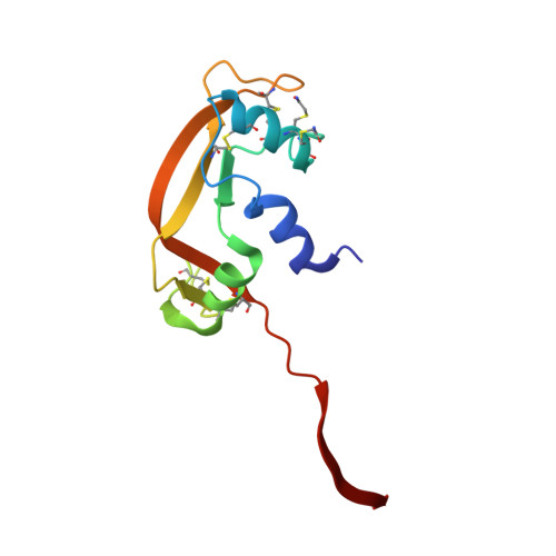
    Bovine seminal ribonuclease (BS-RNase) acquires an interesting anti-tumor activity associated with the swapping on the N-terminal. The first direct experimental evidence on the formation of a C-terminal swapped dimer (C-dimer) obtained from the monomeric derivative of BS-RNase, although under non-native conditions, is here reported. The X-ray model of this dimer reveals a quaternary structure different from that of the C-dimer of RNase A, due to the presence of three mutations in the hinge peptide 111-116. The mutations increase the hinge peptide flexibility and decrease the stability of the C-dimer against dissociation. The biological implications of the structural data are also discussed.


  • Organizational Affiliation
    • Department of Chemical Sciences, University of Naples 'Federico II', via Cintia, 80126 Naples, Italy; Institute of Biostructures and Bioimaging, CNR, Via Mezzocannone 16, 80134 Naples, Italy; National Institute Biostructures and Biosystems, Inter-University Consortium, Viale Medaglie d'Oro 305, I-00136 Rome, Italy.

Macromolecule Content 

  • Total Structure Weight: 27.63 kDa 
  • Atom Count: 1,979 
  • Modeled Residue Count: 248 
  • Deposited Residue Count: 248 
  • Unique protein chains: 1

Macromolecules

Find similar proteins by:|  3D Structure
Entity ID: 1
MoleculeChains  Sequence LengthOrganismDetailsImage
Seminal ribonuclease
A, B
124Bos taurusMutation(s): 4 
Gene Names: SRN
EC: 3.1.27.5 (PDB Primary Data), 4.6.1.18 (UniProt)
UniProt
Find proteins for P00669 (Bos taurus)
Explore P00669 
Go to UniProtKB:  P00669
Entity Groups
Sequence Clusters30% Identity50% Identity70% Identity90% Identity95% Identity100% Identity
UniProt GroupP00669
Sequence Annotations
Expand
Reference Sequence

Small Molecules

Modified Residues  1 Unique
IDChains TypeFormula2D DiagramParent
YCM
Query on YCM
A, B
L-PEPTIDE LINKINGC5 H10 N2 O3 SCYS

Experimental Data & Validation

Experimental Data

  • Method: X-RAY DIFFRACTION
  • Resolution: 2.48 Å
  • R-Value Free:  0.186 (Depositor), 0.196 (DCC) 
  • R-Value Work:  0.170 (Depositor) 
  • R-Value Observed: 0.171 (Depositor) 
Space Group: P 1 21 1
Unit Cell:
Length ( Å )Angle ( ˚ )
a = 28.802α = 90
b = 79.044β = 101.05
c = 75.379γ = 90
Software Package:
Software NamePurpose
HKL-2000data collection
PHASERphasing
PHENIXrefinement
HKL-2000data reduction
HKL-2000data scaling

Structure Validation

View Full Validation Report


Entry History 

Deposition Data

Revision History  (Full details and data files)

  • Version 1.0: 2013-11-06
    Type: Initial release
  • Version 1.1: 2013-12-04
    Changes: Database references
  • Version 1.2: 2023-09-20
    Changes: Data collection, Database references, Derived calculations, Refinement description