4N5S | pdb_00004n5s

Ternary complex structure of Klenow fragment of Taq DNA polymerase I707L mutant (Cs3C KlenTaq) with DNA and ddCTP


Experimental Data Snapshot

  • Method: X-RAY DIFFRACTION
  • Resolution: 1.67 Å
  • R-Value Free: 
    0.192 (Depositor), 0.201 (DCC) 
  • R-Value Work: 
    0.161 (Depositor), 0.171 (DCC) 
  • R-Value Observed: 
    0.163 (Depositor) 

Starting Model: experimental
View more details

wwPDB Validation 3D Report Full Report

Validation slider image for 4N5S

Ligand Structure Quality Assessment 


This is version 1.2 of the entry. See complete history

Literature

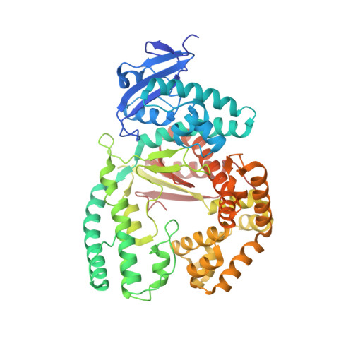
A Conservative Isoleucine to Leucine Mutation Causes Major Rearrangements and Cold Sensitivity in KlenTaq1 DNA Polymerase.

Wu, E.Y.Walsh, A.R.Materne, E.C.Hiltner, E.P.Zielinski, B.Miller, B.R.Mawby, L.Modeste, E.Parish, C.A.Barnes, W.M.Kermekchiev, M.B.

(2015) Biochemistry 54: 881-889

  • DOI: https://doi.org/10.1021/bi501198f
  • Primary Citation Related Structures: 
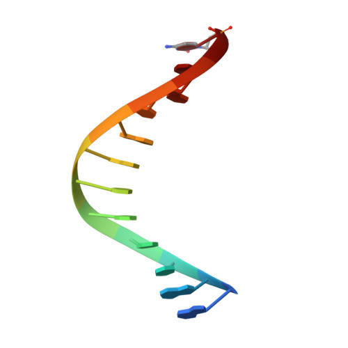
    4N56, 4N5S, 4XIU

  • PubMed Abstract: 

    Assembly of polymerase chain reactions at room temperature can sometimes lead to low yields or unintentional products due to mispriming. Mutation of isoleucine 707 to leucine in DNA polymerase I from Thermus aquaticus substantially decreases its activity at room temperature without compromising its ability to amplify DNA. To understand why a conservative change to the enzyme over 20 Å from the active site can have a large impact on its activity at low temperature, we solved the X-ray crystal structure of the large (5'-to-3' exonuclease-deleted) fragment of Taq DNA polymerase containing the cold-sensitive mutation in the ternary (E-DNA-ddNTP) and binary (E-DNA) complexes. The I707L KlenTaq1 ternary complex was identical to the wild-type in the closed conformation except for the mutation and a rotamer change in nearby phenylalanine 749, suggesting that the enzyme should remain active. However, soaking out of the nucleotide substrate at low temperature results in an altered binary complex made possible by the rotamer change at F749 near the tip of the polymerase O-helix. Surprisingly, two adenosines in the 5'-template overhang fill the vacated active site by stacking with the primer strand, thereby blocking the active site at low temperature. Replacement of the two overhanging adenosines with pyrimidines substantially increased activity at room temperature by keeping the template overhang out of the active site, confirming the importance of base stacking. These results explain the cold-sensitive phenotype of the I707L mutation in KlenTaq1 and serve as an example of a large conformational change affected by a conservative mutation.


  • Organizational Affiliation
    • Department of Biology and ‡Department of Chemistry, University of Richmond , Richmond, Virginia 23173, United States.

Macromolecule Content 

  • Total Structure Weight: 72.27 kDa 
  • Atom Count: 5,394 
  • Modeled Residue Count: 568 
  • Deposited Residue Count: 581 
  • Unique protein chains: 1
  • Unique nucleic acid chains: 2

Macromolecules


Find similar proteins by:|  3D Structure
Entity ID: 3
MoleculeChains  Sequence LengthOrganismDetailsImage
DNA polymerase I, thermostableC [auth A]553Thermus aquaticusMutation(s): 1 
Gene Names: polApol1
EC: 2.7.7.7
UniProt
Find proteins for P19821 (Thermus aquaticus)
Explore P19821 
Go to UniProtKB:  P19821
Entity Groups
Sequence Clusters30% Identity50% Identity70% Identity90% Identity95% Identity100% Identity
UniProt GroupP19821
Sequence Annotations
Expand
Reference Sequence
Find similar nucleic acids by:  Sequence
Entity ID: 1
MoleculeChains LengthOrganismImage
5'-D(*GP*AP*CP*CP*AP*CP*GP*GP*CP*GP*CP*(DOC))-3'A [auth B]12N/A
Sequence Annotations
Expand
Reference Sequence
Find similar nucleic acids by:  Sequence
Entity ID: 2
MoleculeChains LengthOrganismImage
5'-D(*AP*A*AP*GP*GP*GP*CP*GP*CP*CP*GP*TP*GP*GP*TP*C)-3'B [auth C]16N/A
Sequence Annotations
Expand
Reference Sequence

Small Molecules

Ligands 6 Unique
IDChains Name / Formula / InChI Key2D Diagram3D Interactions
DCT

Query on DCT


Download:Ideal Coordinates CCD File
K [auth A]2',3'-DIDEOXYCYTIDINE 5'-TRIPHOSPHATE
C9 H16 N3 O12 P3
ARLKCWCREKRROD-POYBYMJQSA-N
EPE

Query on EPE


Download:Ideal Coordinates CCD File
L [auth A]4-(2-HYDROXYETHYL)-1-PIPERAZINE ETHANESULFONIC ACID
C8 H18 N2 O4 S
JKMHFZQWWAIEOD-UHFFFAOYSA-N
GOL

Query on GOL


Download:Ideal Coordinates CCD File
O [auth A]GLYCEROL
C3 H8 O3
PEDCQBHIVMGVHV-UHFFFAOYSA-N
ACT

Query on ACT


Download:Ideal Coordinates CCD File
M [auth A],
N [auth A]
ACETATE ION
C2 H3 O2
QTBSBXVTEAMEQO-UHFFFAOYSA-M
MN

Query on MN


Download:Ideal Coordinates CCD File
D [auth B]
F [auth A]
G [auth A]
H [auth A]
I [auth A]
D [auth B],
F [auth A],
G [auth A],
H [auth A],
I [auth A],
J [auth A]
MANGANESE (II) ION
Mn
WAEMQWOKJMHJLA-UHFFFAOYSA-N
MG

Query on MG


Download:Ideal Coordinates CCD File
E [auth A]MAGNESIUM ION
Mg
JLVVSXFLKOJNIY-UHFFFAOYSA-N

Experimental Data & Validation

Experimental Data

  • Method: X-RAY DIFFRACTION
  • Resolution: 1.67 Å
  • R-Value Free:  0.192 (Depositor), 0.201 (DCC) 
  • R-Value Work:  0.161 (Depositor), 0.171 (DCC) 
  • R-Value Observed: 0.163 (Depositor) 
Space Group: P 31 2 1
Unit Cell:
Length ( Å )Angle ( ˚ )
a = 107.657α = 90
b = 107.657β = 90
c = 89.659γ = 120
Software Package:
Software NamePurpose
SERGUIdata collection
PHASERphasing
REFMACrefinement
HKL-2000data reduction
HKL-2000data scaling

Structure Validation

View Full Validation Report


Ligand Structure Quality Assessment 


Entry History 

Deposition Data

  • Released Date: 2015-01-21 
  • Deposition Author(s): Wu, E.Y.

Revision History  (Full details and data files)

  • Version 1.0: 2015-01-21
    Type: Initial release
  • Version 1.1: 2015-02-11
    Changes: Database references
  • Version 1.2: 2023-09-20
    Changes: Data collection, Database references, Derived calculations, Refinement description