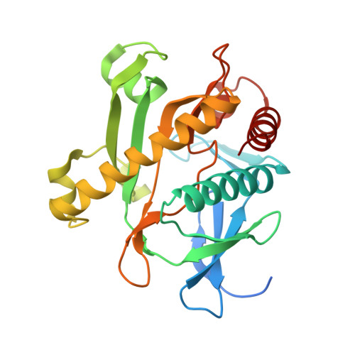
4NYE

Structures of SAICAR Synthetase (PurC) from Streptococcus pneumoniae with ADP, Mg2+, AIR and L-Asp


Experimental Data Snapshot

  • Method: X-RAY DIFFRACTION
  • Resolution: 2.69 Å
  • R-Value Free: 0.315 
  • R-Value Work: 0.228 
  • R-Value Observed: 0.233 

Starting Model: experimental
View more details

wwPDB Validation   3D Report Full Report


Ligand Structure Quality Assessment 


This is version 1.1 of the entry. See complete history


Literature

Structures of SAICAR synthetase (PurC) from Streptococcus pneumoniae with ADP, Mg(2+), AIR and Asp.

Wolf, N.M.Abad-Zapatero, C.Johnson, M.E.Fung, L.W.

(2014) Acta Crystallogr D Biol Crystallogr 70: 841-850

  • DOI: https://doi.org/10.1107/S139900471303366X
  • Primary Citation of Related Structures:  
    4FE2, 4NYE

  • PubMed Abstract: 

    Streptococcus pneumoniae is a multidrug-resistant pathogen that is a target of considerable interest for antibacterial drug development. One strategy for drug discovery is to inhibit an essential metabolic enzyme. The seventh step of the de novo purine-biosynthesis pathway converts carboxyaminoimidazoleribonucleotide (CAIR) and L-aspartic acid (Asp) to 4-(N-succino)-5-aminoimidazole-4-carboxamide ribonucleotide (SAICAR) in the presence of adenosine 5'-triphosphate (ATP) using the enzyme PurC. PurC has been shown to be conditionally essential for bacterial replication. Two crystal structures of this essential enzyme from Streptococcus pneumoniae (spPurC) in the presence of adenosine 5'-diphosphate (ADP), Mg(2+), aminoimidazoleribonucleotide (AIR) and/or Asp have been obtained. This is the first structural study of spPurC, as well as the first of PurC from any species with Asp in the active site. Based on these findings, two model structures are proposed for the active site with all of the essential ligands (ATP, Mg(2+), Asp and CAIR) present, and a relay mechanism for the formation of the product SAICAR is suggested.


  • Organizational Affiliation

    Department of Chemistry, University of Illinois at Chicago, Chicago, IL 60607, USA.


Macromolecules
Find similar proteins by:  (by identity cutoff)  |  3D Structure
Entity ID: 1
MoleculeChains Sequence LengthOrganismDetailsImage
Phosphoribosylaminoimidazole-succinocarboxamide synthase
A, B
255Streptococcus pneumoniae TIGR4Mutation(s): 0 
Gene Names: purCSP_0044
EC: 6.3.2.6
UniProt
Find proteins for Q07296 (Streptococcus pneumoniae serotype 4 (strain ATCC BAA-334 / TIGR4))
Explore Q07296 
Go to UniProtKB:  Q07296
Entity Groups  
Sequence Clusters30% Identity50% Identity70% Identity90% Identity95% Identity100% Identity
UniProt GroupQ07296
Sequence Annotations
Expand
  • Reference Sequence
Small Molecules
Ligands 4 Unique
IDChains Name / Formula / InChI Key2D Diagram3D Interactions
ADP
Query on ADP

Download Ideal Coordinates CCD File 
D [auth A],
H [auth B]
ADENOSINE-5'-DIPHOSPHATE
C10 H15 N5 O10 P2
XTWYTFMLZFPYCI-KQYNXXCUSA-N
ACT
Query on ACT

Download Ideal Coordinates CCD File 
F [auth A],
I [auth B]
ACETATE ION
C2 H3 O2
QTBSBXVTEAMEQO-UHFFFAOYSA-M
CL
Query on CL

Download Ideal Coordinates CCD File 
E [auth A]CHLORIDE ION
Cl
VEXZGXHMUGYJMC-UHFFFAOYSA-M
MG
Query on MG

Download Ideal Coordinates CCD File 
C [auth A],
G [auth B]
MAGNESIUM ION
Mg
JLVVSXFLKOJNIY-UHFFFAOYSA-N
Experimental Data & Validation

Experimental Data

  • Method: X-RAY DIFFRACTION
  • Resolution: 2.69 Å
  • R-Value Free: 0.315 
  • R-Value Work: 0.228 
  • R-Value Observed: 0.233 
  • Space Group: P 1 21 1
Unit Cell:
Length ( Å )Angle ( ˚ )
a = 45.931α = 90
b = 65.111β = 91.96
c = 85.837γ = 90
Software Package:
Software NamePurpose
ADSCdata collection
MOLREPphasing
REFMACrefinement
HKL-2000data reduction
HKL-2000data scaling

Structure Validation

View Full Validation Report


Ligand Structure Quality Assessment 


Entry History 

Deposition Data

Revision History  (Full details and data files)

  • Version 1.0: 2014-04-02
    Type: Initial release
  • Version 1.1: 2023-09-20
    Changes: Data collection, Database references, Derived calculations, Refinement description