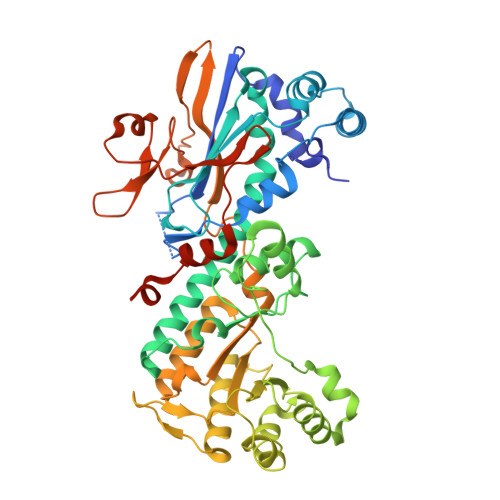
4O19

The crystal structure of a mutant NAMPT (G217V)


Experimental Data Snapshot

  • Method: X-RAY DIFFRACTION
  • Resolution: 1.75 Å
  • R-Value Free: 0.188 
  • R-Value Work: 0.153 
  • R-Value Observed: 0.155 

wwPDB Validation   3D Report Full Report


This is version 1.1 of the entry. See complete history


Literature

Structural Basis for Resistance to Diverse Classes of NAMPT Inhibitors.

Wang, W.Elkins, K.Oh, A.Ho, Y.C.Wu, J.Li, H.Xiao, Y.Kwong, M.Coons, M.Brillantes, B.Cheng, E.Crocker, L.Dragovich, P.S.Sampath, D.Zheng, X.Bair, K.W.O'Brien, T.Belmont, L.D.

(2014) PLoS One 9: e109366-e109366

  • DOI: https://doi.org/10.1371/journal.pone.0109366
  • Primary Citation of Related Structures:  
    4O13, 4O14, 4O15, 4O16, 4O17, 4O18, 4O19, 4O1A, 4O1B, 4O1C, 4O1D, 4O28

  • PubMed Abstract: 

    Inhibiting NAD biosynthesis by blocking the function of nicotinamide phosphoribosyl transferase (NAMPT) is an attractive therapeutic strategy for targeting tumor metabolism. However, the development of drug resistance commonly limits the efficacy of cancer therapeutics. This study identifies mutations in NAMPT that confer resistance to a novel NAMPT inhibitor, GNE-618, in cell culture and in vivo, thus demonstrating that the cytotoxicity of GNE-618 is on target. We determine the crystal structures of six NAMPT mutants in the apo form and in complex with various inhibitors and use cellular, biochemical and structural data to elucidate two resistance mechanisms. One is the surprising finding of allosteric modulation by mutation of residue Ser165, resulting in unwinding of an α-helix that binds the NAMPT substrate 5-phosphoribosyl-1-pyrophosphate (PRPP). The other mechanism is orthosteric blocking of inhibitor binding by mutations of Gly217. Furthermore, by evaluating a panel of diverse small molecule inhibitors, we unravel inhibitor structure activity relationships on the mutant enzymes. These results provide valuable insights into the design of next generation NAMPT inhibitors that offer improved therapeutic potential by evading certain mechanisms of resistance.


  • Organizational Affiliation

    Genentech, Inc., South San Francisco, California, United States of America.


Macromolecules
Find similar proteins by:  (by identity cutoff)  |  3D Structure
Entity ID: 1
MoleculeChains Sequence LengthOrganismDetailsImage
Nicotinamide phosphoribosyltransferase
A, B
501Homo sapiensMutation(s): 1 
Gene Names: NAMPTPBEFPBEF1
EC: 2.4.2.12
UniProt & NIH Common Fund Data Resources
Find proteins for P43490 (Homo sapiens)
Explore P43490 
Go to UniProtKB:  P43490
PHAROS:  P43490
GTEx:  ENSG00000105835 
Entity Groups  
Sequence Clusters30% Identity50% Identity70% Identity90% Identity95% Identity100% Identity
UniProt GroupP43490
Sequence Annotations
Expand
  • Reference Sequence
Small Molecules
Ligands 2 Unique
IDChains Name / Formula / InChI Key2D Diagram3D Interactions
PO4
Query on PO4

Download Ideal Coordinates CCD File 
C [auth A],
G [auth B]
PHOSPHATE ION
O4 P
NBIIXXVUZAFLBC-UHFFFAOYSA-K
EDO
Query on EDO

Download Ideal Coordinates CCD File 
D [auth A]
E [auth A]
F [auth A]
H [auth B]
I [auth B]
D [auth A],
E [auth A],
F [auth A],
H [auth B],
I [auth B],
J [auth B],
K [auth B],
L [auth B],
M [auth B],
N [auth B],
O [auth B]
1,2-ETHANEDIOL
C2 H6 O2
LYCAIKOWRPUZTN-UHFFFAOYSA-N
Experimental Data & Validation

Experimental Data

  • Method: X-RAY DIFFRACTION
  • Resolution: 1.75 Å
  • R-Value Free: 0.188 
  • R-Value Work: 0.153 
  • R-Value Observed: 0.155 
  • Space Group: P 1 21 1
Unit Cell:
Length ( Å )Angle ( ˚ )
a = 60.646α = 90
b = 106.288β = 96.42
c = 83.263γ = 90
Software Package:
Software NamePurpose
B3data collection
PHASERphasing
PHENIXrefinement
HKL-2000data reduction
SCALEPACKdata scaling

Structure Validation

View Full Validation Report


Entry History 

Deposition Data

Revision History  (Full details and data files)

  • Version 1.0: 2014-10-22
    Type: Initial release
  • Version 1.1: 2024-02-28
    Changes: Data collection, Database references, Derived calculations