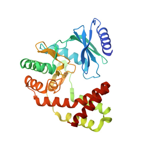
4ORK

Crystal Structure of the Phosphotransferase Domain of the Bifunctional Aminoglycoside Resistance Enzyme AAC(6')-Ie-APH(2'')-Ia


Experimental Data Snapshot

  • Method: X-RAY DIFFRACTION
  • Resolution: 2.30 Å
  • R-Value Free: 0.245 
  • R-Value Work: 0.185 
  • R-Value Observed: 0.188 

wwPDB Validation   3D Report Full Report


Ligand Structure Quality Assessment 


This is version 1.1 of the entry. See complete history


Literature

Structure of the phosphotransferase domain of the bifunctional aminoglycoside-resistance enzyme AAC(6')-Ie-APH(2'')-Ia.

Smith, C.A.Toth, M.Bhattacharya, M.Frase, H.Vakulenko, S.B.

(2014) Acta Crystallogr D Biol Crystallogr 70: 1561-1571

  • DOI: https://doi.org/10.1107/S1399004714005331
  • Primary Citation of Related Structures:  
    4ORK

  • PubMed Abstract: 

    The bifunctional acetyltransferase(6')-Ie-phosphotransferase(2'')-Ia [AAC(6')-Ie-APH(2'')-Ia] is the most important aminoglycoside-resistance enzyme in Gram-positive bacteria, conferring resistance to almost all known aminoglycoside antibiotics in clinical use. Owing to its importance, this enzyme has been the focus of intensive research since its isolation in the mid-1980s but, despite much effort, structural details of AAC(6')-Ie-APH(2'')-Ia have remained elusive. The structure of the Mg2GDP complex of the APH(2'')-Ia domain of the bifunctional enzyme has now been determined at 2.3 Å resolution. The structure of APH(2'')-Ia is reminiscent of the structures of other aminoglycoside phosphotransferases, having a two-domain architecture with the nucleotide-binding site located at the junction of the two domains. Unlike the previously characterized APH(2'')-IIa and APH(2'')-IVa enzymes, which are capable of utilizing both ATP and GTP as the phosphate donors, APH(2'')-Ia uses GTP exclusively in the phosphorylation of the aminoglycoside antibiotics, and in this regard closely resembles the GTP-dependent APH(2'')-IIIa enzyme. In APH(2'')-Ia this GTP selectivity is governed by the presence of a `gatekeeper' residue, Tyr100, the side chain of which projects into the active site and effectively blocks access to the adenine-binding template. Mutation of this tyrosine residue to a less bulky phenylalanine provides better access for ATP to the NTP-binding template and converts APH(2'')-Ia into a dual-specificity enzyme.


  • Organizational Affiliation

    Stanford Synchrotron Radiation Lightsource, Stanford University, Menlo Park, CA 94025, USA.


Macromolecules
Find similar proteins by:  (by identity cutoff)  |  3D Structure
Entity ID: 1
MoleculeChains Sequence LengthOrganismDetailsImage
Bifunctional AAC/APH
A, B, C, D
305Staphylococcus aureusMutation(s): 0 
Gene Names: aacA-aphDR015VRA0030
EC: 2.3.1 (PDB Primary Data), 2.7.1 (PDB Primary Data)
UniProt
Find proteins for P0A0C1 (Staphylococcus aureus)
Explore P0A0C1 
Go to UniProtKB:  P0A0C1
Entity Groups  
Sequence Clusters30% Identity50% Identity70% Identity90% Identity95% Identity100% Identity
UniProt GroupP0A0C1
Sequence Annotations
Expand
  • Reference Sequence
Small Molecules
Ligands 2 Unique
IDChains Name / Formula / InChI Key2D Diagram3D Interactions
GDP
Query on GDP

Download Ideal Coordinates CCD File 
E [auth A],
H [auth B],
K [auth C],
N [auth D]
GUANOSINE-5'-DIPHOSPHATE
C10 H15 N5 O11 P2
QGWNDRXFNXRZMB-UUOKFMHZSA-N
MG
Query on MG

Download Ideal Coordinates CCD File 
F [auth A]
G [auth A]
I [auth B]
J [auth B]
L [auth C]
F [auth A],
G [auth A],
I [auth B],
J [auth B],
L [auth C],
M [auth C],
O [auth D],
P [auth D]
MAGNESIUM ION
Mg
JLVVSXFLKOJNIY-UHFFFAOYSA-N
Experimental Data & Validation

Experimental Data

  • Method: X-RAY DIFFRACTION
  • Resolution: 2.30 Å
  • R-Value Free: 0.245 
  • R-Value Work: 0.185 
  • R-Value Observed: 0.188 
  • Space Group: P 1 21 1
Unit Cell:
Length ( Å )Angle ( ˚ )
a = 90.685α = 90
b = 95.697β = 104.88
c = 91.149γ = 90
Software Package:
Software NamePurpose
Blu-Icedata collection
SOLVEphasing
PHENIXrefinement
XDSdata reduction
SCALAdata scaling

Structure Validation

View Full Validation Report


Ligand Structure Quality Assessment 


Entry History 

Deposition Data

Revision History  (Full details and data files)

  • Version 1.0: 2014-07-02
    Type: Initial release
  • Version 1.1: 2024-02-28
    Changes: Data collection, Database references, Derived calculations