4P1Q | pdb_00004p1q

GREEN FLUORESCENT PROTEIN E222H VARIANT


Experimental Data Snapshot

  • Method: X-RAY DIFFRACTION
  • Resolution: 1.50 Å
  • R-Value Free: 
    0.172 (Depositor), 0.186 (DCC) 
  • R-Value Work: 
    0.136 (Depositor) 
  • R-Value Observed: 
    0.138 (Depositor) 

wwPDB Validation 3D Report Full Report

Validation slider image for 4P1Q

This is version 2.1 of the entry. See complete history

Literature

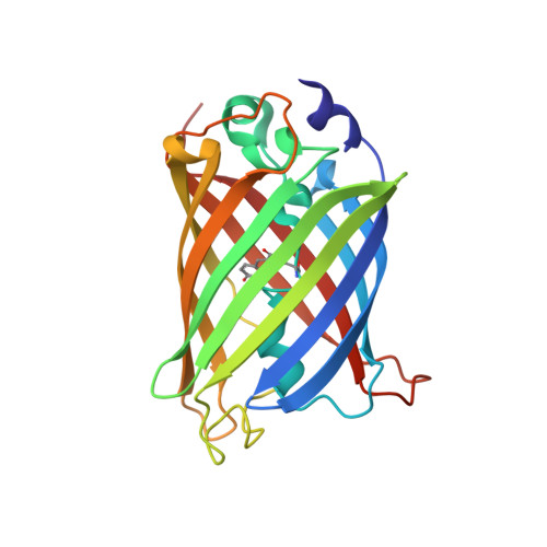
Replacement of Highly Conserved E222 by the Photostable Non-photoconvertible Histidine in GFP.

Auerbach, D.Klein, M.Franz, S.Carius, Y.Lancaster, C.R.Jung, G.

(2014) Chembiochem 15: 1404-1408

  • DOI: https://doi.org/10.1002/cbic.201402075
  • Primary Citation Related Structures: 
    4P1Q

  • PubMed Abstract: 

    The widely used green fluorescent protein (GFP) decarboxylates upon irradiation; this involves removal of the acidic function of the glutamic acid at position 222, thereby resulting in the irreversible photoconversion of GFP. To suppress this phenomenon, the photostable, non-photoconvertible histidine was introduced at position 222 in GFP. The variant E222H shows negligible photodynamic processes and high expression yield. In addition, the stable and bright fluorescence over a wide pH range makes the E222H protein an alternative for GFP in fluorescence imaging and spectroscopy. Other fluorescent proteins are predicted to benefit from replacement of the catalytic glutamic acid by histidine.


  • Organizational Affiliation
    • Department of Biophysical Chemistry, Saarland University, Campus B22, 66123 Saarbrücken (Germany).

Macromolecule Content 

  • Total Structure Weight: 25.85 kDa 
  • Atom Count: 2,181 
  • Modeled Residue Count: 228 
  • Deposited Residue Count: 228 
  • Unique protein chains: 1

Macromolecules

Find similar proteins by:|  3D Structure
Entity ID: 1
MoleculeChains  Sequence LengthOrganismDetailsImage
Green fluorescent protein228Aequorea victoriaMutation(s): 3 
Gene Names: GFP
UniProt
Find proteins for P42212 (Aequorea victoria)
Explore P42212 
Go to UniProtKB:  P42212
Entity Groups
Sequence Clusters30% Identity50% Identity70% Identity90% Identity95% Identity100% Identity
UniProt GroupP42212
Sequence Annotations
Expand
Reference Sequence

Small Molecules

Ligands 1 Unique
IDChains Name / Formula / InChI Key2D Diagram3D Interactions
NA

Query on NA


Download:Ideal Coordinates CCD File
B [auth A]SODIUM ION
Na
FKNQFGJONOIPTF-UHFFFAOYSA-N
Modified Residues  1 Unique
IDChains TypeFormula2D DiagramParent
GYS
Query on GYS
A
L-PEPTIDE LINKINGC14 H15 N3 O5SER, TYR, GLY

Experimental Data & Validation

Experimental Data

  • Method: X-RAY DIFFRACTION
  • Resolution: 1.50 Å
  • R-Value Free:  0.172 (Depositor), 0.186 (DCC) 
  • R-Value Work:  0.136 (Depositor) 
  • R-Value Observed: 0.138 (Depositor) 
Space Group: P 21 21 21
Unit Cell:
Length ( Å )Angle ( ˚ )
a = 50.6α = 90
b = 62.2β = 90
c = 67.57γ = 90
Software Package:
Software NamePurpose
REFMACrefinement

Structure Validation

View Full Validation Report


Entry History 

Deposition Data

Revision History  (Full details and data files)

  • Version 1.0: 2014-07-16
    Type: Initial release
  • Version 1.1: 2015-01-21
    Changes: Structure summary
  • Version 2.0: 2023-11-15
    Changes: Atomic model, Data collection, Database references, Derived calculations, Source and taxonomy
  • Version 2.1: 2024-11-06
    Changes: Structure summary