4P9T | pdb_00004p9t

Structure of the free form of the N-terminal VH1 domain of monomeric alpha-catenin


Experimental Data Snapshot

  • Method: X-RAY DIFFRACTION
  • Resolution: 2.50 Å
  • R-Value Free: 
    0.234 (Depositor), 0.238 (DCC) 
  • R-Value Work: 
    0.215 (Depositor), 0.231 (DCC) 
  • R-Value Observed: 
    0.216 (Depositor) 

Starting Model: experimental
View more details

wwPDB Validation 3D Report Full Report

Validation slider image for 4P9T

This is version 1.4 of the entry. See complete history

Literature

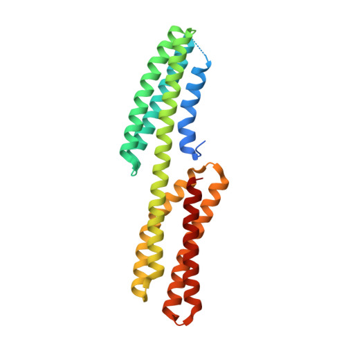
Structure of the free form of the N-terminal VH1 domain of monomeric alpha-catenin.

Shibahara, T.Hirano, Y.Hakoshima, T.

(2015) FEBS Lett 589: 1754-1760

  • DOI: https://doi.org/10.1016/j.febslet.2015.05.053
  • Primary Citation Related Structures: 
    4P9T

  • PubMed Abstract: 

    The N-terminal vinculin-homology 1 (VH1) domain of α-catenin facilitates two exclusive forms, a monomeric form directly bound to β-catenin for linking E-cadherin to F-actin or a homodimer for the inhibition of β-catenin binding. Competition of these two forms is affected by ∼80 N-terminal residues, whose structure is poorly understood. We have determined the structure of the monomeric free form of the αN-catenin VH1 domain and revealed that the N-terminal residues form α1 and α2 helices to complete formation of the N-terminal four-helix bundle. Dynamic conformational changes of these two helices control formation of the β-catenin-bound monomer or unbound homodimer.


  • Organizational Affiliation
    • Structural Biology Laboratory, Nara Institute of Science and Technology, 8916-5 Takayama, Ikoma, Nara 630-0192, Japan.

Macromolecule Content 

  • Total Structure Weight: 119.34 kDa 
  • Atom Count: 6,731 
  • Modeled Residue Count: 924 
  • Deposited Residue Count: 1,052 
  • Unique protein chains: 1

Macromolecules

Find similar proteins by:|  3D Structure
Entity ID: 1
MoleculeChains  Sequence LengthOrganismDetailsImage
Catenin alpha-2
A, B, C, D
263Mus musculusMutation(s): 0 
Gene Names: Ctnna2Catna2
UniProt
Find proteins for Q61301 (Mus musculus)
Explore Q61301 
Go to UniProtKB:  Q61301
Entity Groups
Sequence Clusters30% Identity50% Identity70% Identity90% Identity95% Identity100% Identity
UniProt GroupQ61301
Sequence Annotations
Expand
Reference Sequence

Small Molecules

Ligands 3 Unique
IDChains Name / Formula / InChI Key2D Diagram3D Interactions
IOD

Query on IOD


Download:Ideal Coordinates CCD File
E [auth A]
EA [auth D]
F [auth A]
FA [auth D]
G [auth A]
E [auth A],
EA [auth D],
F [auth A],
FA [auth D],
G [auth A],
GA [auth D],
H [auth A],
HA [auth D],
I [auth A],
IA [auth D],
N [auth B],
O [auth B],
P [auth B],
Q [auth B],
R [auth B],
V [auth C],
W [auth C],
X [auth C],
Y [auth C],
Z [auth C]
IODIDE ION
I
XMBWDFGMSWQBCA-UHFFFAOYSA-M
PEG

Query on PEG


Download:Ideal Coordinates CCD File
DA [auth C],
M [auth A]
DI(HYDROXYETHYL)ETHER
C4 H10 O3
MTHSVFCYNBDYFN-UHFFFAOYSA-N
EDO

Query on EDO


Download:Ideal Coordinates CCD File
AA [auth C]
BA [auth C]
CA [auth C]
J [auth A]
JA [auth D]
AA [auth C],
BA [auth C],
CA [auth C],
J [auth A],
JA [auth D],
K [auth A],
KA [auth D],
L [auth A],
LA [auth D],
S [auth B],
T [auth B],
U [auth B]
1,2-ETHANEDIOL
C2 H6 O2
LYCAIKOWRPUZTN-UHFFFAOYSA-N

Experimental Data & Validation

Experimental Data

  • Method: X-RAY DIFFRACTION
  • Resolution: 2.50 Å
  • R-Value Free:  0.234 (Depositor), 0.238 (DCC) 
  • R-Value Work:  0.215 (Depositor), 0.231 (DCC) 
  • R-Value Observed: 0.216 (Depositor) 
Space Group: P 31
Unit Cell:
Length ( Å )Angle ( ˚ )
a = 68.777α = 90
b = 68.777β = 90
c = 207.965γ = 120
Software Package:
Software NamePurpose
PHENIXrefinement
HKL-2000data scaling
MrBUMPphasing
Cootmodel building

Structure Validation

View Full Validation Report


Entry History 

Deposition Data

Revision History  (Full details and data files)

  • Version 1.0: 2015-04-29
    Type: Initial release
  • Version 1.1: 2015-06-10
    Changes: Database references
  • Version 1.2: 2015-07-15
    Changes: Database references
  • Version 1.3: 2017-11-22
    Changes: Data collection, Database references, Derived calculations, Other, Refinement description, Source and taxonomy, Structure summary
  • Version 1.4: 2023-09-27
    Changes: Data collection, Database references, Refinement description