4PAS | pdb_00004pas

Heterodimeric coiled-coil structure of human GABA(B) receptor


Experimental Data Snapshot

  • Method: X-RAY DIFFRACTION
  • Resolution: 1.62 Å
  • R-Value Free: 
    0.217 (Depositor), 0.217 (DCC) 
  • R-Value Work: 
    0.193 (Depositor), 0.190 (DCC) 
  • R-Value Observed: 
    0.194 (Depositor) 

wwPDB Validation 3D Report Full Report

Validation slider image for 4PAS

This is version 1.5 of the entry. See complete history

Literature

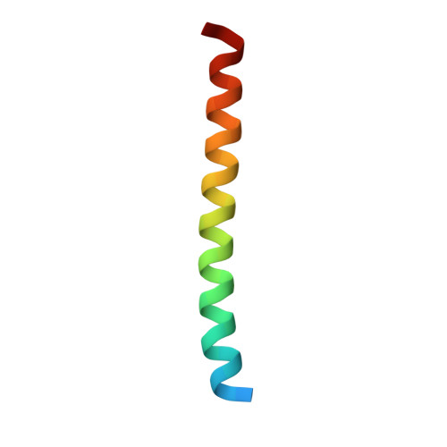
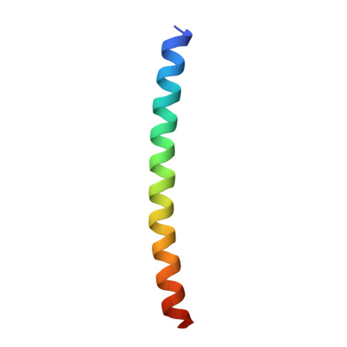
Heterodimeric coiled-coil interactions of human GABAB receptor.

Burmakina, S.Geng, Y.Chen, Y.Fan, Q.R.

(2014) Proc Natl Acad Sci U S A 111: 6958-6963

  • DOI: https://doi.org/10.1073/pnas.1400081111
  • Primary Citation Related Structures: 
    4PAS

  • PubMed Abstract: 

    Metabotropic GABAB receptor is a G protein-coupled receptor that mediates inhibitory neurotransmission in the CNS. It functions as an obligatory heterodimer of GABAB receptor 1 (GBR1) and GABAB receptor 2 (GBR2) subunits. The association between GBR1 and GBR2 masks an endoplasmic reticulum (ER) retention signal in the cytoplasmic region of GBR1 and facilitates cell surface expression of both subunits. Here, we present, to our knowledge, the first crystal structure of an intracellular coiled-coil heterodimer of human GABAB receptor. We found that polar interactions buried within the hydrophobic core determine the specificity of heterodimer pairing. Disruption of the hydrophobic coiled-coil interface with single mutations in either subunit impairs surface expression of GBR1, confirming that the coiled-coil interaction is required to inactivate the adjacent ER retention signal of GBR1. The coiled-coil assembly buries an internalization motif of GBR1 at the heterodimer interface. The ER retention signal of GBR1 is not part of the core coiled-coil structure, suggesting that it is sterically shielded by GBR2 upon heterodimer formation.


  • Organizational Affiliation
    • Departments of Pharmacology and.

Macromolecule Content 

  • Total Structure Weight: 9.74 kDa 
  • Atom Count: 689 
  • Modeled Residue Count: 74 
  • Deposited Residue Count: 82 
  • Unique protein chains: 2

Macromolecules

Find similar proteins by:|  3D Structure
Entity ID: 1
MoleculeChains  Sequence LengthOrganismDetailsImage
Gamma-aminobutyric acid type B receptor subunit 141Homo sapiensMutation(s): 0 
Gene Names: GABBR1GPRC3A
UniProt & NIH Common Fund Data Resources
Find proteins for Q9UBS5 (Homo sapiens)
Explore Q9UBS5 
Go to UniProtKB:  Q9UBS5
PHAROS:  Q9UBS5
GTEx:  ENSG00000204681 
Entity Groups
Sequence Clusters30% Identity50% Identity70% Identity90% Identity95% Identity100% Identity
UniProt GroupQ9UBS5
Sequence Annotations
Expand
Reference Sequence
Find similar proteins by:|  3D Structure
Entity ID: 2
MoleculeChains  Sequence LengthOrganismDetailsImage
Gamma-aminobutyric acid type B receptor subunit 241Homo sapiensMutation(s): 0 
Gene Names: GABBR2GPR51GPRC3B
UniProt & NIH Common Fund Data Resources
Find proteins for O75899 (Homo sapiens)
Explore O75899 
Go to UniProtKB:  O75899
PHAROS:  O75899
GTEx:  ENSG00000136928 
Entity Groups
Sequence Clusters30% Identity50% Identity70% Identity90% Identity95% Identity100% Identity
UniProt GroupO75899
Sequence Annotations
Expand
Reference Sequence

Experimental Data & Validation

Experimental Data

  • Method: X-RAY DIFFRACTION
  • Resolution: 1.62 Å
  • R-Value Free:  0.217 (Depositor), 0.217 (DCC) 
  • R-Value Work:  0.193 (Depositor), 0.190 (DCC) 
  • R-Value Observed: 0.194 (Depositor) 
Space Group: C 2 2 21
Unit Cell:
Length ( Å )Angle ( ˚ )
a = 46.721α = 90
b = 54.654β = 90
c = 55.347γ = 90
Software Package:
Software NamePurpose
HKL-2000data reduction
REFMACrefinement
PDB_EXTRACTdata extraction

Structure Validation

View Full Validation Report


Entry History 

& Funding Information

Deposition Data


Funding OrganizationLocationGrant Number
National Institutes of Health/National Institute of General Medical Sciences (NIH/NIGMS)United States5R01GM088454-05

Revision History  (Full details and data files)

  • Version 1.0: 2014-05-07
    Type: Initial release
  • Version 1.1: 2014-05-14
    Changes: Database references
  • Version 1.2: 2014-07-16
    Changes: Database references
  • Version 1.3: 2017-09-27
    Changes: Author supporting evidence, Database references, Derived calculations, Other, Source and taxonomy
  • Version 1.4: 2019-12-25
    Changes: Author supporting evidence
  • Version 1.5: 2023-12-27
    Changes: Data collection, Database references, Refinement description