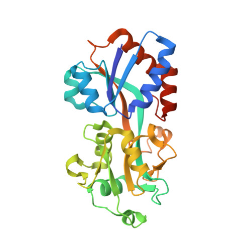
4Q8R | pdb_00004q8r

Crystal structure of a Phosphate Binding Protein (PBP-1) from Clostridium perfringens


Experimental Data Snapshot

  • Method: X-RAY DIFFRACTION
  • Resolution: 1.65 Å
  • R-Value Free: 
    0.178 (Depositor), 0.180 (DCC) 
  • R-Value Work: 
    0.132 (Depositor), 0.142 (DCC) 
  • R-Value Observed: 
    0.135 (Depositor) 

Starting Model: experimental
View more details

wwPDB Validation 3D Report Full Report

Validation slider image for 4Q8R

This is version 1.1 of the entry. See complete history

Literature

Crystal structure of the phosphate-binding protein (PBP-1) of an ABC-type phosphate transporter from Clostridium perfringens.

Gonzalez, D.Richez, M.Bergonzi, C.Chabriere, E.Elias, M.

(2014) Sci Rep 4: 6636-6636

  • DOI: https://doi.org/10.1038/srep06636
  • Primary Citation Related Structures: 
    4Q8R

  • PubMed Abstract: 

    Phosphate limitation is an important environmental stress that affects the metabolism of various organisms and, in particular, can trigger the virulence of numerous bacterial pathogens. Clostridium perfringens, a human pathogen, is one of the most common causes of enteritis necroticans, gas gangrene and food poisoning. Here, we focused on the high affinity phosphate-binding protein (PBP-1) of an ABC-type transporter, responsible for cellular phosphate uptake. We report the crystal structure (1.65 Å resolution) of the protein in complex with phosphate. Interestingly, PBP-1 does not form the short, low-barrier hydrogen bond with phosphate that is typical of previously characterized phosphate-binding proteins, but rather a canonical hydrogen bond. In its unique binding configuration, PBP-1 forms an unusually high number of hydrogen bonds (14) with the phosphate anion. Discrimination experiments reveal that PBP-1 is the least selective PBP characterised so far and is able to discriminate phosphate from its close competing anion, arsenate, by ~150-fold.


  • Organizational Affiliation
    • URMITE UMR CNRS-IRD 6236, IFR48, Faculté de Médecine et de Pharmacie, Université de la Méditerranée, Marseille, France.

Macromolecule Content 

  • Total Structure Weight: 27.64 kDa 
  • Atom Count: 2,097 
  • Modeled Residue Count: 241 
  • Deposited Residue Count: 252 
  • Unique protein chains: 1

Macromolecules

Find similar proteins by:|  3D Structure
Entity ID: 1
MoleculeChains  Sequence LengthOrganismDetailsImage
Phosphate ABC transporter, phosphate-binding protein252Clostridium perfringensMutation(s): 0 
Gene Names: pstSCPF_0617
UniProt
Find proteins for A0A0H2YSI2 (Clostridium perfringens (strain ATCC 13124 / DSM 756 / JCM 1290 / NCIMB 6125 / NCTC 8237 / Type A))
Explore A0A0H2YSI2 
Go to UniProtKB:  A0A0H2YSI2
Entity Groups
Sequence Clusters30% Identity50% Identity70% Identity90% Identity95% Identity100% Identity
UniProt GroupA0A0H2YSI2
Sequence Annotations
Expand
Reference Sequence

Experimental Data & Validation

Experimental Data

  • Method: X-RAY DIFFRACTION
  • Resolution: 1.65 Å
  • R-Value Free:  0.178 (Depositor), 0.180 (DCC) 
  • R-Value Work:  0.132 (Depositor), 0.142 (DCC) 
  • R-Value Observed: 0.135 (Depositor) 
Space Group: F 2 2 2
Unit Cell:
Length ( Å )Angle ( ˚ )
a = 70.78α = 90
b = 105.73β = 90
c = 146.45γ = 90
Software Package:
Software NamePurpose
MOLREPphasing
REFMACrefinement
XDSdata reduction
XSCALEdata scaling

Structure Validation

View Full Validation Report


Entry History 

Deposition Data

Revision History  (Full details and data files)

  • Version 1.0: 2014-11-05
    Type: Initial release
  • Version 1.1: 2023-09-20
    Changes: Data collection, Database references, Derived calculations, Refinement description