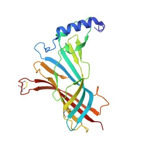
4QAB

X-RAY STRUCTURE of ACETYLCHOLINE BINDING PROTEIN (ACHBP) IN COMPLEX WITH 4-(MORPHOLIN-4-YL)-6-[4-(TRIFLUOROMETHYL)PHENYL]PYRIMIDIN-2-AMINE


Experimental Data Snapshot

  • Method: X-RAY DIFFRACTION
  • Resolution: 2.98 Å
  • R-Value Free: 0.233 
  • R-Value Work: 0.150 
  • R-Value Observed: 0.153 

wwPDB Validation   3D Report Full Report


Ligand Structure Quality Assessment 


This is version 1.5 of the entry. See complete history


Literature

Structural basis for cooperative interactions of substituted 2-aminopyrimidines with the acetylcholine binding protein.

Kaczanowska, K.Harel, M.Radic, Z.Changeux, J.P.Finn, M.G.Taylor, P.

(2014) Proc Natl Acad Sci U S A 111: 10749-10754

  • DOI: https://doi.org/10.1073/pnas.1410992111
  • Primary Citation of Related Structures:  
    4QAA, 4QAB, 4QAC

  • PubMed Abstract: 

    The nicotinic acetylcholine receptor (nAChR) and the acetylcholine binding protein (AChBP) are pentameric oligomers in which binding sites for nicotinic agonists and competitive antagonists are found at selected subunit interfaces. The nAChR spontaneously exists in multiple conformations associated with its activation and desensitization steps, and conformations are selectively stabilized by binding of agonists and antagonists. In the nAChR, agonist binding and the associated conformational changes accompanying activation and desensitization are cooperative. AChBP, which lacks the transmembrane spanning and cytoplasmic domains, serves as a homology model of the extracellular domain of the nAChRs. We identified unique cooperative binding behavior of a number of 4,6-disubstituted 2-aminopyrimidines to Lymnaea AChBP, with different molecular variants exhibiting positive, nH > 1.0, and negative cooperativity, nH < 1.0. Therefore, for a distinctive set of ligands, the extracellular domain of a nAChR surrogate suffices to accommodate cooperative interactions. X-ray crystal structures of AChBP complexes with examples of each allowed the identification of structural features in the ligands that confer differences in cooperative behavior. Both sets of molecules bind at the agonist-antagonist site, as expected from their competition with epibatidine. An analysis of AChBP quaternary structure shows that cooperative ligand binding is associated with a blooming or flare conformation, a structural change not observed with the classical, noncooperative, nicotinic ligands. Positively and negatively cooperative ligands exhibited unique features in the detailed binding determinants and poses of the complexes.


  • Organizational Affiliation

    Department of Pharmacology, Skaggs School of Pharmacy and Pharmaceutical Sciences, University of California, San Diego, La Jolla, CA 92093;


Macromolecules
Find similar proteins by:  (by identity cutoff)  |  3D Structure
Entity ID: 1
MoleculeChains Sequence LengthOrganismDetailsImage
Acetylcholine-binding protein
A, B, C, D, E
A, B, C, D, E, F, G, H, I, J
217Lymnaea stagnalisMutation(s): 0 
UniProt
Find proteins for P58154 (Lymnaea stagnalis)
Explore P58154 
Go to UniProtKB:  P58154
Entity Groups  
Sequence Clusters30% Identity50% Identity70% Identity90% Identity95% Identity100% Identity
UniProt GroupP58154
Glycosylation
Glycosylation Sites: 1
Sequence Annotations
Expand
  • Reference Sequence
Small Molecules
Ligands 3 Unique
IDChains Name / Formula / InChI Key2D Diagram3D Interactions
KK2
Query on KK2

Download Ideal Coordinates CCD File 
BA [auth G]
EA [auth H]
HA [auth I]
K [auth A]
KA [auth J]
BA [auth G],
EA [auth H],
HA [auth I],
K [auth A],
KA [auth J],
N [auth B],
P [auth C],
S [auth D],
V [auth E],
Z [auth F]
4-(morpholin-4-yl)-6-[4-(trifluoromethyl)phenyl]pyrimidin-2-amine
C15 H15 F3 N4 O
XZLXJMNYYMKDDT-UHFFFAOYSA-N
NAG
Query on NAG

Download Ideal Coordinates CCD File 
CA [auth G]
FA [auth H]
IA [auth I]
L [auth A]
LA [auth J]
CA [auth G],
FA [auth H],
IA [auth I],
L [auth A],
LA [auth J],
O [auth B],
Q [auth C],
T [auth D],
W [auth E]
2-acetamido-2-deoxy-beta-D-glucopyranose
C8 H15 N O6
OVRNDRQMDRJTHS-FMDGEEDCSA-N
PO4
Query on PO4

Download Ideal Coordinates CCD File 
AA [auth F]
DA [auth G]
GA [auth H]
JA [auth I]
M [auth A]
AA [auth F],
DA [auth G],
GA [auth H],
JA [auth I],
M [auth A],
MA [auth J],
R [auth C],
U [auth D],
X [auth E],
Y [auth E]
PHOSPHATE ION
O4 P
NBIIXXVUZAFLBC-UHFFFAOYSA-K
Experimental Data & Validation

Experimental Data

  • Method: X-RAY DIFFRACTION
  • Resolution: 2.98 Å
  • R-Value Free: 0.233 
  • R-Value Work: 0.150 
  • R-Value Observed: 0.153 
  • Space Group: P 21 21 21
Unit Cell:
Length ( Å )Angle ( ˚ )
a = 122.639α = 90
b = 134.015β = 90
c = 147.02γ = 90
Software Package:
Software NamePurpose
HKL-2000data collection
PHASERphasing
PHENIXrefinement
HKL-2000data reduction
HKL-2000data scaling

Structure Validation

View Full Validation Report


Ligand Structure Quality Assessment 


Entry History 

Deposition Data

Revision History  (Full details and data files)

  • Version 1.0: 2014-07-16
    Type: Initial release
  • Version 1.1: 2014-07-30
    Changes: Database references
  • Version 1.2: 2014-08-13
    Changes: Database references
  • Version 1.3: 2018-01-24
    Changes: Structure summary
  • Version 1.4: 2020-07-29
    Type: Remediation
    Reason: Carbohydrate remediation
    Changes: Data collection, Database references, Derived calculations, Structure summary
  • Version 1.5: 2024-10-30
    Changes: Data collection, Database references, Structure summary