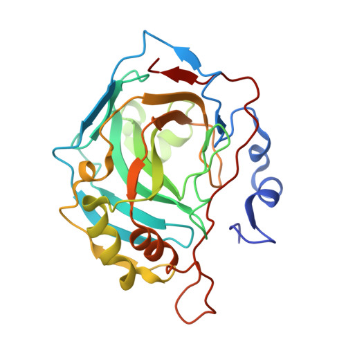
4QSB

Crystal structure of human carbonic anhydrase isozyme II with 3-{[(4-methyl-6-oxo-1,6-dihydropyrimidin-2-yl)thio]acetyl}benzenesulfonamide


Experimental Data Snapshot

  • Method: X-RAY DIFFRACTION
  • Resolution: 1.40 Å
  • R-Value Free: 0.220 
  • R-Value Work: 0.188 
  • R-Value Observed: 0.192 

Starting Model: experimental
View more details

wwPDB Validation   3D Report Full Report


Ligand Structure Quality Assessment 


This is version 1.1 of the entry. See complete history


Literature

Intrinsic Thermodynamics and Structure Correlation of Benzenesulfonamides with a Pyrimidine Moiety Binding to Carbonic Anhydrases I, II, VII, XII, and XIII

Kisonaite, M.Zubriene, A.Capkauskaite, E.Smirnov, A.Smirnoviene, J.Kairys, V.Michailoviene, V.Manakova, E.Grazulis, S.Matulis, D.

(2014) PLoS One 9: e114106-e114106

  • DOI: https://doi.org/10.1371/journal.pone.0114106
  • Primary Citation of Related Structures:  
    4QSA, 4QSB, 4QSI, 4QSJ

  • PubMed Abstract: 

    The early stage of drug discovery is often based on selecting the highest affinity lead compound. To this end the structural and energetic characterization of the binding reaction is important. The binding energetics can be resolved into enthalpic and entropic contributions to the binding Gibbs free energy. Most compound binding reactions are coupled to the absorption or release of protons by the protein or the compound. A distinction between the observed and intrinsic parameters of the binding energetics requires the dissection of the protonation/deprotonation processes. Since only the intrinsic parameters can be correlated with molecular structural perturbations associated with complex formation, it is these parameters that are required for rational drug design. Carbonic anhydrase (CA) isoforms are important therapeutic targets to treat a range of disorders including glaucoma, obesity, epilepsy, and cancer. For effective treatment isoform-specific inhibitors are needed. In this work we investigated the binding and protonation energetics of sixteen [(2-pyrimidinylthio)acetyl]benzenesulfonamide CA inhibitors using isothermal titration calorimetry and fluorescent thermal shift assay. The compounds were built by combining four sulfonamide headgroups with four tailgroups yielding 16 compounds. Their intrinsic binding thermodynamics showed the limitations of the functional group energetic additivity approach used in fragment-based drug design, especially at the level of enthalpies and entropies of binding. Combined with high resolution crystal structural data correlations were drawn between the chemical functional groups on selected inhibitors and intrinsic thermodynamic parameters of CA-inhibitor complex formation.


  • Organizational Affiliation

    Department of Biothermodynamics and Drug Design, Institute of Biotechnology, Vilnius University, Graičiūno 8, Vilnius, LT-02241, Lithuania.


Macromolecules
Find similar proteins by:  (by identity cutoff)  |  3D Structure
Entity ID: 1
MoleculeChains Sequence LengthOrganismDetailsImage
Carbonic anhydrase 2260Homo sapiensMutation(s): 0 
Gene Names: CA2
EC: 4.2.1.1 (PDB Primary Data), 4.2.1.69 (UniProt)
UniProt & NIH Common Fund Data Resources
Find proteins for P00918 (Homo sapiens)
Explore P00918 
Go to UniProtKB:  P00918
PHAROS:  P00918
GTEx:  ENSG00000104267 
Entity Groups  
Sequence Clusters30% Identity50% Identity70% Identity90% Identity95% Identity100% Identity
UniProt GroupP00918
Sequence Annotations
Expand
  • Reference Sequence
Small Molecules
Ligands 4 Unique
IDChains Name / Formula / InChI Key2D Diagram3D Interactions
EWY
Query on EWY

Download Ideal Coordinates CCD File 
C [auth A]3-{[(4-methyl-6-oxo-1,6-dihydropyrimidin-2-yl)sulfanyl]acetyl}benzenesulfonamide
C13 H13 N3 O4 S2
NYRNFJQJOXIJHL-UHFFFAOYSA-N
MES
Query on MES

Download Ideal Coordinates CCD File 
D [auth A]2-(N-MORPHOLINO)-ETHANESULFONIC ACID
C6 H13 N O4 S
SXGZJKUKBWWHRA-UHFFFAOYSA-N
DMS
Query on DMS

Download Ideal Coordinates CCD File 
E [auth A],
F [auth A]
DIMETHYL SULFOXIDE
C2 H6 O S
IAZDPXIOMUYVGZ-UHFFFAOYSA-N
ZN
Query on ZN

Download Ideal Coordinates CCD File 
B [auth A]ZINC ION
Zn
PTFCDOFLOPIGGS-UHFFFAOYSA-N
Experimental Data & Validation

Experimental Data

  • Method: X-RAY DIFFRACTION
  • Resolution: 1.40 Å
  • R-Value Free: 0.220 
  • R-Value Work: 0.188 
  • R-Value Observed: 0.192 
  • Space Group: P 1 21 1
Unit Cell:
Length ( Å )Angle ( ˚ )
a = 42.247α = 90
b = 40.984β = 104.04
c = 71.48γ = 90
Software Package:
Software NamePurpose
REFMACrefinement
MOSFLMdata reduction
SCALAdata scaling
PDB_EXTRACTdata extraction
MAR345dtbdata collection
MOLREPphasing

Structure Validation

View Full Validation Report


Ligand Structure Quality Assessment 


Entry History 

Deposition Data

Revision History  (Full details and data files)

  • Version 1.0: 2015-01-21
    Type: Initial release
  • Version 1.1: 2023-11-08
    Changes: Data collection, Database references, Derived calculations, Refinement description