4RCV | pdb_00004rcv

M. tuberculosis 1-deoxy-d-xylulose-5-phosphate reductoisomerase bound to 1-deoxy-L-erythrulose


Experimental Data Snapshot

  • Method: X-RAY DIFFRACTION
  • Resolution: 2.29 Å
  • R-Value Free: 
    0.232 (Depositor), 0.234 (DCC) 
  • R-Value Work: 
    0.173 (Depositor), 0.174 (DCC) 
  • R-Value Observed: 
    0.176 (Depositor) 

Starting Model: experimental
View more details

wwPDB Validation 3D Report Full Report

Validation slider image for 4RCV

Ligand Structure Quality Assessment 


This is version 1.1 of the entry. See complete history

Literature

The role of phosphate in a multistep enzymatic reaction: reactions of the substrate and intermediate in pieces.

Kholodar, S.A.Allen, C.L.Gulick, A.M.Murkin, A.S.

(2015) J Am Chem Soc 137: 2748-2756

  • DOI: https://doi.org/10.1021/ja512911f
  • Primary Citation Related Structures: 
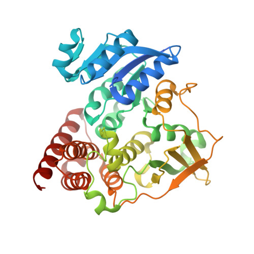
    4RCV

  • PubMed Abstract: 

    Several mechanistically unrelated enzymes utilize the binding energy of their substrate's nonreacting phosphoryl group to accelerate catalysis. Evidence for the involvement of the phosphodianion in transition state formation has come from reactions of the substrate in pieces, in which reaction of a truncated substrate lacking its phosphorylmethyl group is activated by inorganic phosphite. What has remained unknown until now is how the phosphodianion group influences the reaction energetics at different points along the reaction coordinate. 1-Deoxy-D-xylulose-5-phosphate (DXP) reductoisomerase (DXR), which catalyzes the isomerization of DXP to 2-C-methyl-D-erythrose 4-phosphate (MEsP) and subsequent NADPH-dependent reduction, presents a unique opportunity to address this concern. Previously, we have reported the effect of covalently linked phosphate on the energetics of DXP turnover. Through the use of chemically synthesized MEsP and its phosphate-truncated analogue, 2-C-methyl-D-glyceraldehyde, the current study revealed a loss of 6.1 kcal/mol of kinetic barrier stabilization upon truncation, of which 4.4 kcal/mol was regained in the presence of phosphite dianion. The activating effect of phosphite was accompanied by apparent tightening of its interactions within the active site at the intermediate stage of the reaction, suggesting a role of the phosphodianion in disfavoring intermediate release and in modulation of the on-enzyme isomerization equilibrium. The results of kinetic isotope effect and structural studies indicate rate limitation by physical steps when the covalent linkage is severed. These striking differences in the energetics of the natural reaction and the reactions in pieces provide a deeper insight into the contribution of enzyme-phosphodianion interactions to the reaction coordinate.


  • Organizational Affiliation
    • Department of Chemistry, University at Buffalo , Buffalo, New York 14260-3000, United States.

Macromolecule Content 

  • Total Structure Weight: 86.8 kDa 
  • Atom Count: 5,666 
  • Modeled Residue Count: 747 
  • Deposited Residue Count: 808 
  • Unique protein chains: 1

Macromolecules

Find similar proteins by:|  3D Structure
Entity ID: 1
MoleculeChains  Sequence LengthOrganismDetailsImage
1-deoxy-D-xylulose 5-phosphate reductoisomerase
A, B
404Mycobacterium tuberculosis H37RvMutation(s): 0 
Gene Names: dxrP425_02990RVBD_2870c
EC: 1.1.1.267
UniProt
Find proteins for P9WNS1 (Mycobacterium tuberculosis (strain ATCC 25618 / H37Rv))
Explore P9WNS1 
Go to UniProtKB:  P9WNS1
Entity Groups
Sequence Clusters30% Identity50% Identity70% Identity90% Identity95% Identity100% Identity
UniProt GroupP9WNS1
Sequence Annotations
Expand
Reference Sequence

Small Molecules

Ligands 4 Unique
IDChains Name / Formula / InChI Key2D Diagram3D Interactions
NDP

Query on NDP


Download:Ideal Coordinates CCD File
D [auth A],
H [auth B]
NADPH DIHYDRO-NICOTINAMIDE-ADENINE-DINUCLEOTIDE PHOSPHATE
C21 H30 N7 O17 P3
ACFIXJIJDZMPPO-NNYOXOHSSA-N
DE2

Query on DE2


Download:Ideal Coordinates CCD File
E [auth A]1-deoxy-L-erythrulose
C4 H8 O3
SEYLPRWNVFCVRQ-BYPYZUCNSA-N
PO3

Query on PO3


Download:Ideal Coordinates CCD File
F [auth A],
I [auth B]
PHOSPHITE ION
O3 P
AQSJGOWTSHOLKH-UHFFFAOYSA-N
MN

Query on MN


Download:Ideal Coordinates CCD File
C [auth A],
G [auth B]
MANGANESE (II) ION
Mn
WAEMQWOKJMHJLA-UHFFFAOYSA-N

Experimental Data & Validation

Experimental Data

  • Method: X-RAY DIFFRACTION
  • Resolution: 2.29 Å
  • R-Value Free:  0.232 (Depositor), 0.234 (DCC) 
  • R-Value Work:  0.173 (Depositor), 0.174 (DCC) 
  • R-Value Observed: 0.176 (Depositor) 
Space Group: P 1 21 1
Unit Cell:
Length ( Å )Angle ( ˚ )
a = 67.37α = 90
b = 64.378β = 101.73
c = 85.741γ = 90
Software Package:
Software NamePurpose
Blu-Icedata collection
PHENIXmodel building
PHASERphasing
PHENIXrefinement
HKL-2000data reduction
HKL-2000data scaling
PHENIXphasing

Structure Validation

View Full Validation Report


Ligand Structure Quality Assessment 


Entry History 

Deposition Data

Revision History  (Full details and data files)

  • Version 1.0: 2015-03-18
    Type: Initial release
  • Version 1.1: 2023-09-20
    Changes: Data collection, Database references, Derived calculations, Refinement description