4RTB | pdb_00004rtb

X-ray structure of the FeFe-hydrogenase maturase HydG from Carboxydothermus hydrogenoformans


Experimental Data Snapshot

  • Method: X-RAY DIFFRACTION
  • Resolution: 2.79 Å
  • R-Value Free: 
    0.255 (Depositor), 0.257 (DCC) 
  • R-Value Work: 
    0.223 (Depositor), 0.232 (DCC) 
  • R-Value Observed: 
    0.225 (Depositor) 

Starting Model: experimental
View more details

wwPDB Validation 3D Report Full Report

Validation slider image for 4RTB

Ligand Structure Quality Assessment 


This is version 1.2 of the entry. See complete history

Literature

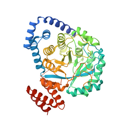
Crystal Structure of HydG from Carboxydothermus hydrogenoformans: A Trifunctional [FeFe]-Hydrogenase Maturase.

Nicolet, Y.Pagnier, A.Zeppieri, L.Martin, L.Amara, P.Fontecilla-Camps, J.C.

(2015) Chembiochem 16: 397-402

  • DOI: https://doi.org/10.1002/cbic.201402661
  • Primary Citation Related Structures: 
    4RTB

  • PubMed Abstract: 

    The structure of the radical S-adenosyl-L-methionine (SAM) [FeFe]-hydrogenase maturase HydG involved in CN(-) /CO synthesis is characterized by two internal tunnels connecting its tyrosine-binding pocket with the external medium and the C-terminal Fe4 S4 cluster-containing region. A comparison with a tryptophan-bound NosL structure suggests that substrate binding causes the closing of the first tunnel and, along with mutagenesis studies, that tyrosine binds to HydG with its amino group well positioned for H-abstraction by SAM. In this orientation the dehydroglycine (DHG) fragment caused by tyrosine Cα-Cβ bond scission can readily migrate through the second tunnel towards the C-terminal domain where both CN(-) and CO are synthesized. Our HydG structure appears to be in a relaxed state with its C-terminal cluster CysX2 CysX22 Cys motif exposed to solvent. A rotation of this domain coupled to Fe4 S4 cluster assembly would bury its putatively reactive unique Fe ion thereby allowing it to interact with DHG.


  • Organizational Affiliation
    • Metalloproteins Unit, Institut de Biologie Structurale, UMR5075, CEA, CNRS, Université Grenoble Alpes, 71, Avenue des Martyrs, CS 10090, 38044 Grenoble cedex 9 (France). yvain.nicolet@ibs.fr.

Macromolecule Content 

  • Total Structure Weight: 55.65 kDa 
  • Atom Count: 3,367 
  • Modeled Residue Count: 429 
  • Deposited Residue Count: 477 
  • Unique protein chains: 1

Macromolecules

Find similar proteins by:|  3D Structure
Entity ID: 1
MoleculeChains  Sequence LengthOrganismDetailsImage
HydG protein477Carboxydothermus hydrogenoformans Z-2901Mutation(s): 0 
Gene Names: CHY_1549
UniProt
Find proteins for Q3ABV3 (Carboxydothermus hydrogenoformans (strain ATCC BAA-161 / DSM 6008 / Z-2901))
Explore Q3ABV3 
Go to UniProtKB:  Q3ABV3
Entity Groups
Sequence Clusters30% Identity50% Identity70% Identity90% Identity95% Identity100% Identity
UniProt GroupQ3ABV3
Sequence Annotations
Expand
Reference Sequence

Small Molecules

Ligands 3 Unique
IDChains Name / Formula / InChI Key2D Diagram3D Interactions
SAM

Query on SAM


Download:Ideal Coordinates CCD File
B [auth A]S-ADENOSYLMETHIONINE
C15 H22 N6 O5 S
MEFKEPWMEQBLKI-FCKMPRQPSA-N
SF4

Query on SF4


Download:Ideal Coordinates CCD File
C [auth A]IRON/SULFUR CLUSTER
Fe4 S4
LJBDFODJNLIPKO-UHFFFAOYSA-N
CL

Query on CL


Download:Ideal Coordinates CCD File
D [auth A],
E [auth A],
F [auth A],
G [auth A],
H [auth A]
CHLORIDE ION
Cl
VEXZGXHMUGYJMC-UHFFFAOYSA-M

Experimental Data & Validation

Experimental Data

  • Method: X-RAY DIFFRACTION
  • Resolution: 2.79 Å
  • R-Value Free:  0.255 (Depositor), 0.257 (DCC) 
  • R-Value Work:  0.223 (Depositor), 0.232 (DCC) 
  • R-Value Observed: 0.225 (Depositor) 
Space Group: P 41 21 2
Unit Cell:
Length ( Å )Angle ( ˚ )
a = 92.23α = 90
b = 92.23β = 90
c = 167.6γ = 90
Software Package:
Software NamePurpose
ADSCdata collection
PHASERphasing
PHENIXrefinement
XDSdata reduction
XDSdata scaling

Structure Validation

View Full Validation Report


Ligand Structure Quality Assessment 


Entry History 

Deposition Data

Revision History  (Full details and data files)

  • Version 1.0: 2015-01-28
    Type: Initial release
  • Version 1.1: 2015-02-25
    Changes: Database references
  • Version 1.2: 2023-09-20
    Changes: Data collection, Database references, Derived calculations, Refinement description