4S0N

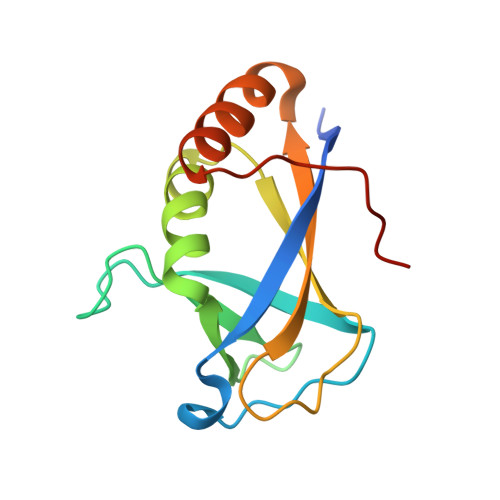
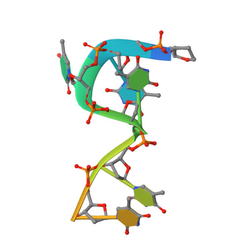
Crystal Structure of HLTF HIRAN Domain bound to DNA


Experimental Data Snapshot

  • Method: X-RAY DIFFRACTION
  • Resolution: 1.50 Å
  • R-Value Free: 0.185 
  • R-Value Work: 0.153 
  • R-Value Observed: 0.154 

wwPDB Validation   3D Report Full Report


Ligand Structure Quality Assessment 


This is version 1.2 of the entry. See complete history


Literature

HLTF's Ancient HIRAN Domain Binds 3' DNA Ends to Drive Replication Fork Reversal.

Kile, A.C.Chavez, D.A.Bacal, J.Eldirany, S.Korzhnev, D.M.Bezsonova, I.Eichman, B.F.Cimprich, K.A.

(2015) Mol Cell 58: 1090-1100

  • DOI: https://doi.org/10.1016/j.molcel.2015.05.013
  • Primary Citation of Related Structures:  
    2MZN, 4S0N

  • PubMed Abstract: 

    Stalled replication forks are a critical problem for the cell because they can lead to complex genome rearrangements that underlie cell death and disease. Processes such as DNA damage tolerance and replication fork reversal protect stalled forks from these events. A central mediator of these DNA damage responses in humans is the Rad5-related DNA translocase, HLTF. Here, we present biochemical and structural evidence that the HIRAN domain, an ancient and conserved domain found in HLTF and other DNA processing proteins, is a modified oligonucleotide/oligosaccharide (OB) fold that binds to 3' ssDNA ends. We demonstrate that the HIRAN domain promotes HLTF-dependent fork reversal in vitro through its interaction with 3' ssDNA ends found at forks. Finally, we show that HLTF restrains replication fork progression in cells in a HIRAN-dependent manner. These findings establish a mechanism of HLTF-mediated fork reversal and provide insight into the requirement for distinct fork remodeling activities in the cell.


  • Organizational Affiliation

    Department of Chemical and Systems Biology, Stanford University School of Medicine, Stanford, CA 94305, USA.


Macromolecules

Find similar proteins by:  (by identity cutoff)  |  3D Structure
Entity ID: 1
MoleculeChains Sequence LengthOrganismDetailsImage
Helicase-like transcription factor
A, B, C, D
130Homo sapiensMutation(s): 0 
Gene Names: HLTFHIP116ARNF80SMARCA3SNF2L3ZBU1
EC: 3.6.4 (PDB Primary Data), 6.3.2 (PDB Primary Data), 2.3.2.27 (UniProt)
UniProt & NIH Common Fund Data Resources
Find proteins for Q14527 (Homo sapiens)
Explore Q14527 
Go to UniProtKB:  Q14527
PHAROS:  Q14527
GTEx:  ENSG00000071794 
Entity Groups  
Sequence Clusters30% Identity50% Identity70% Identity90% Identity95% Identity100% Identity
UniProt GroupQ14527
Sequence Annotations
Expand
  • Reference Sequence

Find similar nucleic acids by:  Sequence   |   3D Structure  

Entity ID: 2
MoleculeChains LengthOrganismImage
5'-D(*TP*TP*TP*TP*TP*TP*TP*TP*TP*T)-3'
E, F, G, H
10synthetic construct
Sequence Annotations
Expand
  • Reference Sequence
Small Molecules
Ligands 3 Unique
IDChains Name / Formula / InChI Key2D Diagram3D Interactions
DT
Query on DT

Download Ideal Coordinates CCD File 
I [auth A],
J [auth A]
THYMIDINE-5'-MONOPHOSPHATE
C10 H15 N2 O8 P
GYOZYWVXFNDGLU-XLPZGREQSA-N
GOL
Query on GOL

Download Ideal Coordinates CCD File 
K [auth A],
M [auth B]
GLYCEROL
C3 H8 O3
PEDCQBHIVMGVHV-UHFFFAOYSA-N
NA
Query on NA

Download Ideal Coordinates CCD File 
L [auth A],
N [auth B],
O [auth C],
P [auth D]
SODIUM ION
Na
FKNQFGJONOIPTF-UHFFFAOYSA-N
Experimental Data & Validation

Experimental Data

Unit Cell:
Length ( Å )Angle ( ˚ )
a = 61.026α = 90
b = 74.211β = 113.68
c = 66.18γ = 90
Software Package:
Software NamePurpose
MAR345dtbdata collection
SHARPphasing
PHENIXrefinement
HKL-2000data reduction
HKL-2000data scaling

Structure Validation

View Full Validation Report


Ligand Structure Quality Assessment 


Entry History 

Deposition Data

Revision History  (Full details and data files)

  • Version 1.0: 2015-05-27
    Type: Initial release
  • Version 1.1: 2015-07-01
    Changes: Database references
  • Version 1.2: 2024-02-28
    Changes: Data collection, Database references, Derived calculations