4UM9

Crystal structure of alpha V beta 6 with peptide


Experimental Data Snapshot

  • Method: X-RAY DIFFRACTION
  • Resolution: 2.50 Å
  • R-Value Free: 
    0.266 (Depositor), 0.270 (DCC) 
  • R-Value Work: 
    0.222 (Depositor), 0.230 (DCC) 
  • R-Value Observed: 
    0.223 (Depositor) 

Starting Model: experimental
View more details

wwPDB Validation   3D Report Full Report


Ligand Structure Quality Assessment 

Created with Raphaël 2.3.0Worse 01 BetterLigand structure goodness of fit to experimental dataBest fitted NAGClick on this verticalbar to view detailsBest fitted MANClick on this verticalbar to view details

This is version 3.2 of the entry. See complete history


Literature

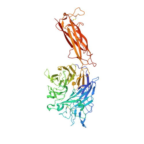
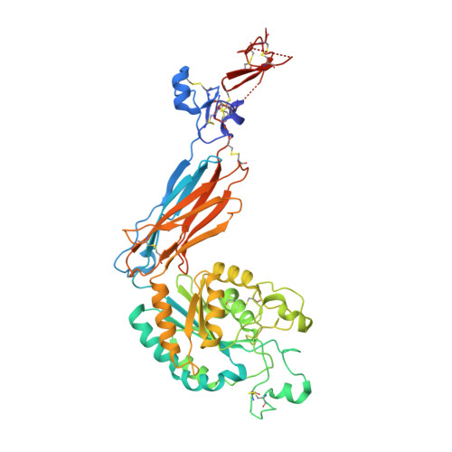
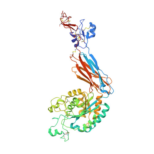
Structural Determinants of Integrin Beta-Subunit Specificity for Latent Tgf-Beta

Dong, X.Hudson, N.E.Lu, C.Springer, T.A.

(2014) Nat Struct Mol Biol 21: 1091

  • DOI: https://doi.org/10.1038/nsmb.2905
  • Primary Citation of Related Structures:  
    4UM8, 4UM9

  • PubMed Abstract: 

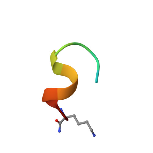
    Eight integrin α-β heterodimers recognize ligands with an Arg-Gly-Asp (RGD) motif. However, the structural mechanism by which integrins differentiate among extracellular proteins with RGD motifs is not understood. Here, crystal structures, mutations and peptide-affinity measurements show that αVβ6 binds with high affinity to a RGDLXXL/I motif within the prodomains of TGF-β1 and TGF-β3. The LXXL/I motif forms an amphipathic α-helix that binds in a hydrophobic pocket in the β6 subunit. Elucidation of the basis for ligand binding specificity by the integrin β subunit reveals contributions by three different βI-domain loops, which we designate specificity-determining loops (SDLs) 1, 2 and 3. Variation in a pair of single key residues in SDL1 and SDL3 correlates with the variation of the entire β subunit in integrin evolution, thus suggesting a paradigmatic role in overall β-subunit function.


  • Organizational Affiliation

    1] Boston Children's Hospital, Harvard Medical School, Boston, Massachusetts, USA. [2] Department of Biological Chemistry and Molecular Pharmacology, Harvard Medical School, Boston, Massachusetts, USA.


Macromolecules
Find similar proteins by:  (by identity cutoff)  |  3D Structure
Entity ID: 1
MoleculeChains Sequence LengthOrganismDetailsImage
Integrin alpha-V heavy chain
A, C
604Homo sapiensMutation(s): 0 
Gene Names: ITGAVMSK8VNRAVTNR
UniProt & NIH Common Fund Data Resources
Find proteins for P06756 (Homo sapiens)
Explore P06756 
Go to UniProtKB:  P06756
PHAROS:  P06756
GTEx:  ENSG00000138448 
Entity Groups  
Sequence Clusters30% Identity50% Identity70% Identity90% Identity95% Identity100% Identity
UniProt GroupP06756
Glycosylation
Glycosylation Sites: 6Go to GlyGen: P06756-1
Sequence Annotations
Expand
  • Reference Sequence
Find similar proteins by:  (by identity cutoff)  |  3D Structure
Entity ID: 2
MoleculeChains Sequence LengthOrganismDetailsImage
Integrin beta-6483Homo sapiensMutation(s): 0 
Gene Names: ITGB6
UniProt & NIH Common Fund Data Resources
Find proteins for P18564 (Homo sapiens)
Explore P18564 
Go to UniProtKB:  P18564
PHAROS:  P18564
GTEx:  ENSG00000115221 
Entity Groups  
Sequence Clusters30% Identity50% Identity70% Identity90% Identity95% Identity100% Identity
UniProt GroupP18564
Glycosylation
Glycosylation Sites: 3Go to GlyGen: P18564-1
Sequence Annotations
Expand
  • Reference Sequence
Find similar proteins by:  (by identity cutoff)  |  3D Structure
Entity ID: 3
MoleculeChains Sequence LengthOrganismDetailsImage
Integrin beta-6483Homo sapiensMutation(s): 0 
Gene Names: ITGB6
UniProt & NIH Common Fund Data Resources
Find proteins for P18564 (Homo sapiens)
Explore P18564 
Go to UniProtKB:  P18564
PHAROS:  P18564
GTEx:  ENSG00000115221 
Entity Groups  
Sequence Clusters30% Identity50% Identity70% Identity90% Identity95% Identity100% Identity
UniProt GroupP18564
Glycosylation
Glycosylation Sites: 3Go to GlyGen: P18564-1
Sequence Annotations
Expand
  • Reference Sequence

Find similar proteins by:  Sequence   |   3D Structure  

Entity ID: 4
MoleculeChains Sequence LengthOrganismDetailsImage
Latency-associated peptide
E, F
13Homo sapiensMutation(s): 0 
Gene Names: TGFB3
UniProt & NIH Common Fund Data Resources
Find proteins for P10600 (Homo sapiens)
Explore P10600 
Go to UniProtKB:  P10600
PHAROS:  P10600
GTEx:  ENSG00000119699 
Entity Groups  
Sequence Clusters30% Identity50% Identity70% Identity90% Identity95% Identity100% Identity
UniProt GroupP10600
Sequence Annotations
Expand
  • Reference Sequence
Oligosaccharides

Help

Entity ID: 5
MoleculeChains Length2D Diagram Glycosylation3D Interactions
2-acetamido-2-deoxy-beta-D-glucopyranose-(1-4)-2-acetamido-2-deoxy-beta-D-glucopyranose
G, J, K, L, M
2N-Glycosylation
Glycosylation Resources
GlyTouCan:  G42666HT
GlyCosmos:  G42666HT
GlyGen:  G42666HT
Entity ID: 6
MoleculeChains Length2D Diagram Glycosylation3D Interactions
alpha-D-mannopyranose-(1-6)-beta-D-mannopyranose-(1-4)-2-acetamido-2-deoxy-beta-D-glucopyranose-(1-4)-2-acetamido-2-deoxy-beta-D-glucopyranose
H
4N-Glycosylation
Glycosylation Resources
GlyTouCan:  G22573RC
GlyCosmos:  G22573RC
GlyGen:  G22573RC
Entity ID: 7
MoleculeChains Length2D Diagram Glycosylation3D Interactions
alpha-D-mannopyranose-(1-3)-alpha-D-mannopyranose-(1-6)-[alpha-D-mannopyranose-(1-3)]beta-D-mannopyranose-(1-4)-2-acetamido-2-deoxy-beta-D-glucopyranose-(1-4)-2-acetamido-2-deoxy-beta-D-glucopyranose
I, N
6N-Glycosylation
Glycosylation Resources
GlyTouCan:  G09724ZC
GlyCosmos:  G09724ZC
GlyGen:  G09724ZC
Entity ID: 8
MoleculeChains Length2D Diagram Glycosylation3D Interactions
beta-D-mannopyranose-(1-4)-2-acetamido-2-deoxy-beta-D-glucopyranose-(1-4)-2-acetamido-2-deoxy-beta-D-glucopyranose
O
3N-Glycosylation
Glycosylation Resources
GlyTouCan:  G15407YE
GlyCosmos:  G15407YE
GlyGen:  G15407YE
Small Molecules
Ligands 5 Unique
IDChains Name / Formula / InChI Key2D Diagram3D Interactions
NAG
Query on NAG

Download Ideal Coordinates CCD File 
BA [auth B]
CA [auth B]
DA [auth B]
KA [auth C]
MA [auth C]
2-acetamido-2-deoxy-beta-D-glucopyranose
C8 H15 N O6
OVRNDRQMDRJTHS-FMDGEEDCSA-N
MAN
Query on MAN

Download Ideal Coordinates CCD File 
LA [auth C]alpha-D-mannopyranose
C6 H12 O6
WQZGKKKJIJFFOK-PQMKYFCFSA-N
SO4
Query on SO4

Download Ideal Coordinates CCD File 
EA [auth C]
FA [auth C]
Q [auth A]
R [auth A]
S [auth A]
SULFATE ION
O4 S
QAOWNCQODCNURD-UHFFFAOYSA-L
CA
Query on CA

Download Ideal Coordinates CCD File 
AA [auth B]
GA [auth C]
HA [auth C]
IA [auth C]
JA [auth C]
CALCIUM ION
Ca
BHPQYMZQTOCNFJ-UHFFFAOYSA-N
MG
Query on MG

Download Ideal Coordinates CCD File 
NA [auth D],
Y [auth B]
MAGNESIUM ION
Mg
JLVVSXFLKOJNIY-UHFFFAOYSA-N
Experimental Data & Validation

Experimental Data

  • Method: X-RAY DIFFRACTION
  • Resolution: 2.50 Å
  • R-Value Free:  0.266 (Depositor), 0.270 (DCC) 
  • R-Value Work:  0.222 (Depositor), 0.230 (DCC) 
  • R-Value Observed: 0.223 (Depositor) 
Space Group: C 1 2 1
Unit Cell:
Length ( Å )Angle ( ˚ )
a = 185.02α = 90
b = 168.09β = 98.95
c = 101.8γ = 90
Software Package:
Software NamePurpose
PHENIXrefinement
XDSdata reduction
XDSdata scaling
PHASERphasing

Structure Validation

View Full Validation Report


Ligand Structure Quality Assessment 

Created with Raphaël 2.3.0Worse 01 BetterLigand structure goodness of fit to experimental dataBest fitted NAGClick on this verticalbar to view detailsBest fitted MANClick on this verticalbar to view details

Entry History 

Deposition Data

Revision History  (Full details and data files)

  • Version 1.0: 2014-11-12
    Type: Initial release
  • Version 1.1: 2014-11-19
    Changes: Database references
  • Version 1.2: 2014-12-17
    Changes: Database references
  • Version 1.3: 2016-03-16
    Changes: Database references, Derived calculations, Other, Structure summary
  • Version 2.0: 2020-07-29
    Type: Remediation
    Reason: Carbohydrate remediation
    Changes: Advisory, Atomic model, Data collection, Derived calculations, Other, Structure summary
  • Version 3.0: 2023-03-01
    Changes: Advisory, Atomic model, Data collection, Database references, Derived calculations, Non-polymer description, Polymer sequence, Source and taxonomy, Structure summary
  • Version 3.1: 2024-02-07
    Changes: Data collection, Refinement description
  • Version 3.2: 2024-11-06
    Changes: Structure summary