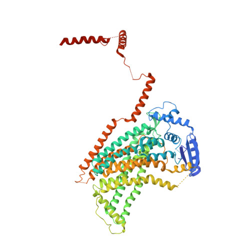
4WIT

TMEM16 lipid scramblase in crystal form 2


Experimental Data Snapshot

  • Method: X-RAY DIFFRACTION
  • Resolution: 3.40 Å
  • R-Value Free: 0.292 
  • R-Value Work: 0.248 
  • R-Value Observed: 0.250 

wwPDB Validation   3D Report Full Report


This is version 1.3 of the entry. See complete history


Literature

X-ray structure of a calcium-activated TMEM16 lipid scramblase.

Brunner, J.D.Lim, N.K.Schenck, S.Duerst, A.Dutzler, R.

(2014) Nature 516: 207-212

  • DOI: https://doi.org/10.1038/nature13984
  • Primary Citation of Related Structures:  
    4WIS, 4WIT

  • PubMed Abstract: 

    The TMEM16 family of proteins, also known as anoctamins, features a remarkable functional diversity. This family contains the long sought-after Ca(2+)-activated chloride channels as well as lipid scramblases and cation channels. Here we present the crystal structure of a TMEM16 family member from the fungus Nectria haematococca that operates as a Ca(2+)-activated lipid scramblase. Each subunit of the homodimeric protein contains ten transmembrane helices and a hydrophilic membrane-traversing cavity that is exposed to the lipid bilayer as a potential site of catalysis. This cavity harbours a conserved Ca(2+)-binding site located within the hydrophobic core of the membrane. Mutations of residues involved in Ca(2+) coordination affect both lipid scrambling in N. haematococca TMEM16 and ion conduction in the Cl(-) channel TMEM16A. The structure reveals the general architecture of the family and its mode of Ca(2+) activation. It also provides insight into potential scrambling mechanisms and serves as a framework to unravel the conduction of ions in certain TMEM16 proteins.


  • Organizational Affiliation

    Department of Biochemistry, University of Zurich, Winterthurerstrasse 190, CH-8057 Zurich, Switzerland.


Macromolecules
Find similar proteins by:  (by identity cutoff)  |  3D Structure
Entity ID: 1
MoleculeChains Sequence LengthOrganismDetailsImage
Predicted protein
A, B
735Fusarium vanettenii 77-13-4Mutation(s): 0 
Gene Names: NECHADRAFT_66456
Membrane Entity: Yes 
UniProt
Find proteins for C7Z7K1 (Fusarium vanettenii (strain ATCC MYA-4622 / CBS 123669 / FGSC 9596 / NRRL 45880 / 77-13-4))
Explore C7Z7K1 
Go to UniProtKB:  C7Z7K1
Entity Groups  
Sequence Clusters30% Identity50% Identity70% Identity90% Identity95% Identity100% Identity
UniProt GroupC7Z7K1
Sequence Annotations
Expand
  • Reference Sequence
Experimental Data & Validation

Experimental Data

  • Method: X-RAY DIFFRACTION
  • Resolution: 3.40 Å
  • R-Value Free: 0.292 
  • R-Value Work: 0.248 
  • R-Value Observed: 0.250 
  • Space Group: P 21 21 21
Unit Cell:
Length ( Å )Angle ( ˚ )
a = 115.94α = 90
b = 127.24β = 90
c = 180.11γ = 90
Software Package:
Software NamePurpose
PHENIXrefinement
XDSdata reduction
SHELXphasing
SHARPphasing
DMphasing
Omodel building
Cootmodel building
SCALEPACKdata scaling

Structure Validation

View Full Validation Report


Entry History & Funding Information

Deposition Data


Funding OrganizationLocationGrant Number
European Research CouncilSwitzerland339116-AnoBest
SNSFSwitzerlandNCCR TransCure

Revision History  (Full details and data files)

  • Version 1.0: 2014-11-12
    Type: Initial release
  • Version 1.1: 2014-11-19
    Changes: Database references
  • Version 1.2: 2014-12-17
    Changes: Database references
  • Version 1.3: 2024-05-08
    Changes: Author supporting evidence, Data collection, Database references, Derived calculations