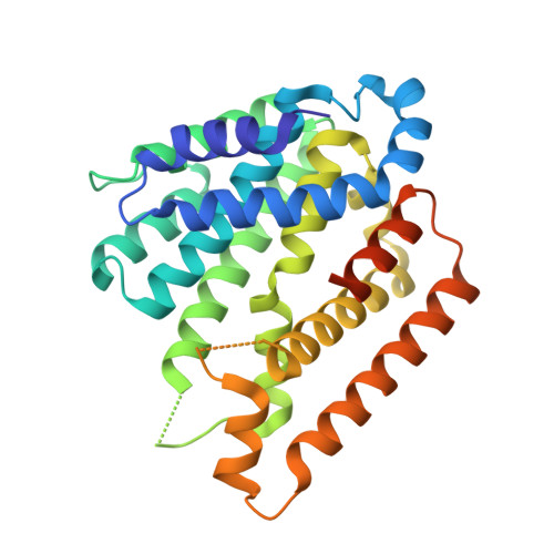
4XLY

The complex structure of KS-D75C with substrate CPP


Experimental Data Snapshot

  • Method: X-RAY DIFFRACTION
  • Resolution: 1.82 Å
  • R-Value Free: 0.240 
  • R-Value Work: 0.200 

Starting Model: experimental
View more details

wwPDB Validation   3D Report Full Report


Ligand Structure Quality Assessment 


This is version 1.1 of the entry. See complete history


Literature

Structure, function and inhibition of ent-kaurene synthase from Bradyrhizobium japonicum.

Liu, W.Feng, X.Zheng, Y.Huang, C.H.Nakano, C.Hoshino, T.Bogue, S.Ko, T.P.Chen, C.C.Cui, Y.Li, J.Wang, I.Hsu, S.T.Oldfield, E.Guo, R.T.

(2014) Sci Rep 4: 6214

  • DOI: https://doi.org/10.1038/srep06214
  • Primary Citation of Related Structures:  
    4W4R, 4W4S, 4XLX, 4XLY

  • PubMed Abstract: 

    We report the first X-ray crystal structure of ent-kaur-16-ene synthase from Bradyrhizobium japonicum, together with the results of a site-directed mutagenesis investigation into catalytic activity. The structure is very similar to that of the α domains of modern plant terpene cyclases, a result that is of interest since it has been proposed that many plant terpene cyclases may have arisen from bacterial diterpene cyclases. The ent-copalyl diphosphate substrate binds to a hydrophobic pocket near a cluster of Asp and Arg residues that are essential for catalysis, with the carbocations formed on ionization being protected by Leu, Tyr and Phe residues. A bisphosphonate inhibitor binds to the same site. In the kaurene synthase from the moss Physcomitrella patens, 16-α-hydroxy-ent-kaurane as well as kaurene are produced since Leu and Tyr in the P. patens kaurene synthase active site are replaced by smaller residues enabling carbocation quenching by water. Overall, the results represent the first structure determination of a bacterial diterpene cyclase, providing insights into catalytic activity, as well as structural comparisons with diverse terpene synthases and cyclases which clearly separate the terpene cyclases from other terpene synthases having highly α-helical structures.


  • Organizational Affiliation

    1] Industrial Enzymes National Engineering Laboratory, Tianjin Institute of Industrial Biotechnology, Chinese Academy of Sciences, Tianjin 300308, China [2].


Macromolecules
Find similar proteins by:  (by identity cutoff)  |  3D Structure
Entity ID: 1
MoleculeChains Sequence LengthOrganismDetailsImage
Uncharacterized protein blr2150
A, B
300Bradyrhizobium diazoefficiens USDA 110Mutation(s): 1 
Gene Names: blr2150
UniProt
Find proteins for Q45222 (Bradyrhizobium diazoefficiens (strain JCM 10833 / BCRC 13528 / IAM 13628 / NBRC 14792 / USDA 110))
Explore Q45222 
Go to UniProtKB:  Q45222
Entity Groups  
Sequence Clusters30% Identity50% Identity70% Identity90% Identity95% Identity100% Identity
UniProt GroupQ45222
Sequence Annotations
Expand
  • Reference Sequence
Small Molecules
Ligands 1 Unique
IDChains Name / Formula / InChI Key2D Diagram3D Interactions
ECP
Query on ECP

Download Ideal Coordinates CCD File 
C [auth A],
D [auth B]
(2E)-3-methyl-5-[(1R,4aR,8aR)-5,5,8a-trimethyl-2-methylidenedecahydronaphthalen-1-yl]pent-2-en-1-yl trihydrogen diphosphate
C20 H36 O7 P2
JCAIWDXKLCEQEO-PGHZQYBFSA-N
Experimental Data & Validation

Experimental Data

  • Method: X-RAY DIFFRACTION
  • Resolution: 1.82 Å
  • R-Value Free: 0.240 
  • R-Value Work: 0.200 
  • Space Group: P 41
Unit Cell:
Length ( Å )Angle ( ˚ )
a = 65.814α = 90
b = 65.814β = 90
c = 136.244γ = 90
Software Package:
Software NamePurpose
HKL-2000data collection
PHASERmodel building
CNSrefinement
HKL-2000data reduction
HKL-2000data scaling
PHASERphasing

Structure Validation

View Full Validation Report


Ligand Structure Quality Assessment 


Entry History 

Deposition Data

Revision History  (Full details and data files)

  • Version 1.0: 2015-02-04
    Type: Initial release
  • Version 1.1: 2023-11-08
    Changes: Data collection, Database references, Derived calculations, Refinement description, Source and taxonomy