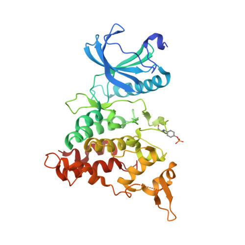
4YLL

Crystal structure of DYRK1AA in complex with 10-Bromo-substituted 11H-indolo[3,2-c]quinolone-6-carboxylic acid inhibitor 5t


Experimental Data Snapshot

  • Method: X-RAY DIFFRACTION
  • Resolution: 1.40 Å
  • R-Value Free: 0.172 
  • R-Value Work: 0.151 
  • R-Value Observed: 0.152 

Starting Model: experimental
View more details

wwPDB Validation   3D Report Full Report


Ligand Structure Quality Assessment 


This is version 1.5 of the entry. See complete history


Literature

10-Iodo-11H-indolo[3,2-c]quinoline-6-carboxylic Acids Are Selective Inhibitors of DYRK1A.

Falke, H.Chaikuad, A.Becker, A.Loaec, N.Lozach, O.Abu Jhaisha, S.Becker, W.Jones, P.G.Preu, L.Baumann, K.Knapp, S.Meijer, L.Kunick, C.

(2015) J Med Chem 58: 3131-3143

  • DOI: https://doi.org/10.1021/jm501994d
  • Primary Citation of Related Structures:  
    4YLJ, 4YLK, 4YLL

  • PubMed Abstract: 

    The protein kinase DYRK1A has been suggested to act as one of the intracellular regulators contributing to neurological alterations found in individuals with Down syndrome. For an assessment of the role of DYRK1A, selective synthetic inhibitors are valuable pharmacological tools. However, the DYRK1A inhibitors described in the literature so far either are not sufficiently selective or have not been tested against closely related kinases from the DYRK and the CLK protein kinase families. The aim of this study was the identification of DYRK1A inhibitors exhibiting selectivity versus the structurally and functionally closely related DYRK and CLK isoforms. Structure modification of the screening hit 11H-indolo[3,2-c]quinoline-6-carboxylic acid revealed structure-activity relationships for kinase inhibition and enabled the design of 10-iodo-substituted derivatives as very potent DYRK1A inhibitors with considerable selectivity against CLKs. X-ray structure determination of three 11H-indolo[3,2-c]quinoline-6-carboxylic acids cocrystallized with DYRK1A confirmed the predicted binding mode within the ATP binding site.


  • Organizational Affiliation

    †Institut für Medizinische und Pharmazeutische Chemie, Technische Universität Braunschweig, Beethovenstraße 55, 38106 Braunschweig, Germany.


Macromolecules
Find similar proteins by:  (by identity cutoff)  |  3D Structure
Entity ID: 1
MoleculeChains Sequence LengthOrganismDetailsImage
Dual specificity tyrosine-phosphorylation-regulated kinase 1A361Homo sapiensMutation(s): 0 
Gene Names: DYRK1ADYRKMNBMNBH
EC: 2.7.12.1 (PDB Primary Data), 2.7.11.23 (UniProt)
UniProt & NIH Common Fund Data Resources
Find proteins for Q13627 (Homo sapiens)
Explore Q13627 
Go to UniProtKB:  Q13627
PHAROS:  Q13627
GTEx:  ENSG00000157540 
Entity Groups  
Sequence Clusters30% Identity50% Identity70% Identity90% Identity95% Identity100% Identity
UniProt GroupQ13627
Sequence Annotations
Expand
  • Reference Sequence
Small Molecules
Ligands 3 Unique
IDChains Name / Formula / InChI Key2D Diagram3D Interactions
4E3
Query on 4E3

Download Ideal Coordinates CCD File 
P [auth A]10-bromo-2-iodo-11H-indolo[3,2-c]quinoline-6-carboxylic acid
C16 H8 Br I N2 O2
WYQPGNJIFZHADC-UHFFFAOYSA-N
SO4
Query on SO4

Download Ideal Coordinates CCD File 
B [auth A],
C [auth A],
D [auth A],
E [auth A],
F [auth A]
SULFATE ION
O4 S
QAOWNCQODCNURD-UHFFFAOYSA-L
EDO
Query on EDO

Download Ideal Coordinates CCD File 
G [auth A]
H [auth A]
I [auth A]
J [auth A]
K [auth A]
G [auth A],
H [auth A],
I [auth A],
J [auth A],
K [auth A],
L [auth A],
M [auth A],
N [auth A],
O [auth A]
1,2-ETHANEDIOL
C2 H6 O2
LYCAIKOWRPUZTN-UHFFFAOYSA-N
Modified Residues  1 Unique
IDChains TypeFormula2D DiagramParent
PTR
Query on PTR
A
L-PEPTIDE LINKINGC9 H12 N O6 PTYR
Experimental Data & Validation

Experimental Data

  • Method: X-RAY DIFFRACTION
  • Resolution: 1.40 Å
  • R-Value Free: 0.172 
  • R-Value Work: 0.151 
  • R-Value Observed: 0.152 
  • Space Group: C 1 2 1
Unit Cell:
Length ( Å )Angle ( ˚ )
a = 100.03α = 90
b = 69.86β = 117.72
c = 67.9γ = 90
Software Package:
Software NamePurpose
REFMACrefinement
MOSFLMdata reduction
SCALAdata scaling
PHASERphasing

Structure Validation

View Full Validation Report


Ligand Structure Quality Assessment 


Entry History 

Revision History  (Full details and data files)

  • Version 1.0: 2015-03-25
    Type: Initial release
  • Version 1.1: 2015-04-22
    Changes: Database references
  • Version 1.2: 2017-11-22
    Changes: Data collection, Database references, Derived calculations, Refinement description, Source and taxonomy
  • Version 1.3: 2023-09-27
    Changes: Data collection, Database references, Refinement description
  • Version 1.4: 2023-11-15
    Changes: Data collection
  • Version 1.5: 2024-11-13
    Changes: Structure summary