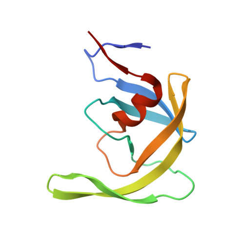
4Z4X

Crystal Structure of Multidrug Resistant HIV-1 Protease Clinical Isolate PR20D25N with Open Flap


Experimental Data Snapshot

  • Method: X-RAY DIFFRACTION
  • Resolution: 1.75 Å
  • R-Value Free: 0.217 
  • R-Value Work: 0.184 
  • R-Value Observed: 0.186 

Starting Model: experimental
View more details

wwPDB Validation   3D Report Full Report


This is version 1.3 of the entry. See complete history


Literature

Conformational variation of an extreme drug resistant mutant of HIV protease.

Shen, C.H.Chang, Y.C.Agniswamy, J.Harrison, R.W.Weber, I.T.

(2015) J Mol Graph Model 62: 87-96

  • DOI: https://doi.org/10.1016/j.jmgm.2015.09.006
  • Primary Citation of Related Structures:  
    4Z4X, 4Z50

  • PubMed Abstract: 

    Molecular mechanisms leading to high level drug resistance have been analyzed for the clinical variant of HIV-1 protease bearing 20 mutations (PR20); which has several orders of magnitude worse affinity for tested drugs. Two crystal structures of ligand-free PR20 with the D25N mutation of the catalytic aspartate (PR20D25N) revealed three dimers with different flap conformations. The diverse conformations of PR20D25N included a dimer with one flap in a unique "tucked" conformation; directed into the active site. Analysis of molecular dynamics (MD) simulations of the ligand-free PR20 and wild-type enzymes showed that the mutations in PR20 alter the correlated interactions between two monomers in the dimer. The two flaps tend to fluctuate more independently in PR20 than in the wild type enzyme. Combining the results of structural analysis by X-ray crystallography and MD simulations; unusual flap conformations and weakly correlated inter-subunit motions may contribute to the high level resistance of PR20.


  • Organizational Affiliation

    Department of Biology, Georgia State University, Atlanta, GA 30303, USA.


Macromolecules
Find similar proteins by:  (by identity cutoff)  |  3D Structure
Entity ID: 1
MoleculeChains Sequence LengthOrganismDetailsImage
Protease
A, B
99Human immunodeficiency virus 1Mutation(s): 12 
Gene Names: pol
UniProt
Find proteins for Q0PQ60 (Human immunodeficiency virus 1)
Explore Q0PQ60 
Go to UniProtKB:  Q0PQ60
Entity Groups  
Sequence Clusters30% Identity50% Identity70% Identity90% Identity95% Identity100% Identity
UniProt GroupQ0PQ60
Sequence Annotations
Expand
  • Reference Sequence
Experimental Data & Validation

Experimental Data

  • Method: X-RAY DIFFRACTION
  • Resolution: 1.75 Å
  • R-Value Free: 0.217 
  • R-Value Work: 0.184 
  • R-Value Observed: 0.186 
  • Space Group: P 41
Unit Cell:
Length ( Å )Angle ( ˚ )
a = 45.522α = 90
b = 45.522β = 90
c = 104.148γ = 90
Software Package:
Software NamePurpose
REFMACrefinement
MOLREPphasing
HKL-2000data scaling
SERGUIdata collection
HKL-2000data reduction

Structure Validation

View Full Validation Report


Entry History & Funding Information

Deposition Data


Funding OrganizationLocationGrant Number
National Institutes of Health/National Institute of General Medical Sciences (NIH/NIGMS)United StatesU01GM062920

Revision History  (Full details and data files)

  • Version 1.0: 2015-10-14
    Type: Initial release
  • Version 1.1: 2017-09-27
    Changes: Author supporting evidence, Derived calculations
  • Version 1.2: 2019-12-25
    Changes: Author supporting evidence
  • Version 1.3: 2023-09-27
    Changes: Data collection, Database references, Refinement description