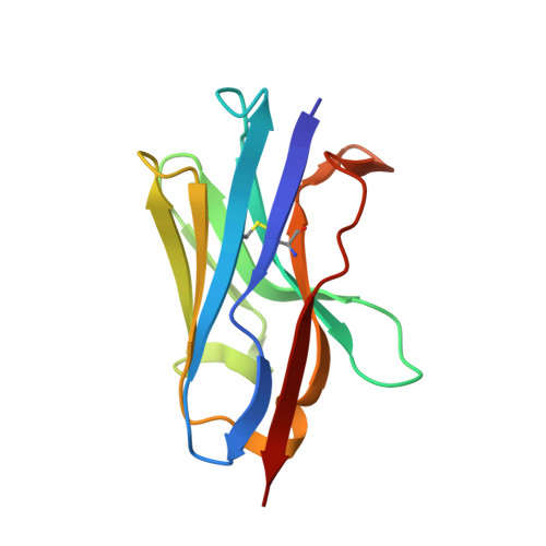
4ZG1 | pdb_00004zg1

Crystal structure of a nanobody raised against KDM5B


Experimental Data Snapshot

  • Method: X-RAY DIFFRACTION
  • Resolution: 1.85 Å
  • R-Value Free: 
    0.235 (Depositor), 0.235 (DCC) 
  • R-Value Work: 
    0.186 (Depositor), 0.188 (DCC) 
  • R-Value Observed: 
    0.188 (Depositor) 

Starting Model: experimental
View more details

wwPDB Validation 3D Report Full Report

Validation slider image for 4ZG1

This is version 1.6 of the entry. See complete history

Literature

Structure and binding properties of a cameloid nanobody raised against KDM5B.

Wiuf, A.Kristensen, L.H.Kristensen, O.Dorosz, J.Jensen, J.Gajhede, M.

(2015) Acta Crystallogr F Struct Biol Commun 71: 1235-1241

  • DOI: https://doi.org/10.1107/S2053230X1501537X
  • Primary Citation Related Structures: 
    4ZG1

  • PubMed Abstract: 

    The histone demethylase KDM5B is considered to be a promising target for anticancer therapy. Single-chain antibodies from llama (nanobodies) have been raised to aid in crystallization and structure determination of this enzyme. The antigen-binding properties of 15 of these nanobodies have been characterized. The crystal structure of one of these (NB17) has been determined to a resolution of 1.85 Å. NB17 crystallizes in space group P4322 with six molecules in the asymmetric unit. The six molecules in the asymmetric unit pack as an entity with approximate D3 symmetry with interactions mediated by the CDR loops, which could act as a crystallization nucleus. NB17 does not bind to monomeric KDM5B residues 1-820, but is found to bind to aggregates formed after incubation at 310 K.


  • Organizational Affiliation
    • Department of Drug Design and Pharmacology, University of Copenhagen, 2200 Copenhagen, Denmark.

Macromolecule Content 

  • Total Structure Weight: 74.88 kDa 
  • Atom Count: 6,181 
  • Modeled Residue Count: 701 
  • Deposited Residue Count: 702 
  • Unique protein chains: 1

Macromolecules

Find similar proteins by:|  3D Structure
Entity ID: 1
MoleculeChains  Sequence LengthOrganismDetailsImage
NB17
A, B, C, D, E
A, B, C, D, E, F
117Lama glamaMutation(s): 0 

Experimental Data & Validation

Experimental Data

  • Method: X-RAY DIFFRACTION
  • Resolution: 1.85 Å
  • R-Value Free:  0.235 (Depositor), 0.235 (DCC) 
  • R-Value Work:  0.186 (Depositor), 0.188 (DCC) 
  • R-Value Observed: 0.188 (Depositor) 
Space Group: P 43 2 2
Unit Cell:
Length ( Å )Angle ( ˚ )
a = 91.52α = 90
b = 91.52β = 90
c = 184.59γ = 90
Software Package:
Software NamePurpose
PHENIXrefinement
SCALAdata scaling
PDB_EXTRACTdata extraction
PHASERphasing
XDSdata reduction

Structure Validation

View Full Validation Report


Entry History 

Deposition Data

Revision History  (Full details and data files)

  • Version 1.0: 2015-05-06
    Type: Initial release
  • Version 1.1: 2015-10-21
    Changes: Database references
  • Version 1.2: 2018-01-17
    Changes: Data collection
  • Version 1.3: 2019-10-09
    Changes: Data collection
  • Version 1.4: 2019-10-16
    Changes: Data collection
  • Version 1.5: 2024-01-10
    Changes: Data collection, Database references, Refinement description
  • Version 1.6: 2024-10-23
    Changes: Structure summary