4ZG4 | pdb_00004zg4

Myosin Vc Pre-powerstroke


Experimental Data Snapshot

  • Method: X-RAY DIFFRACTION
  • Resolution: 2.36 Å
  • R-Value Free: 
    0.213 (Depositor), 0.220 (DCC) 
  • R-Value Work: 
    0.173 (Depositor), 0.184 (DCC) 
  • R-Value Observed: 
    0.175 (Depositor) 

Starting Model: experimental
View more details

wwPDB Validation 3D Report Full Report

Validation slider image for 4ZG4

Ligand Structure Quality Assessment 


This is version 1.4 of the entry. See complete history

Literature

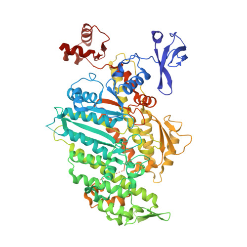
Force-producing ADP state of myosin bound to actin.

Wulf, S.F.Ropars, V.Fujita-Becker, S.Oster, M.Hofhaus, G.Trabuco, L.G.Pylypenko, O.Sweeney, H.L.Houdusse, A.M.Schroder, R.R.

(2016) Proc Natl Acad Sci U S A 113: E1844-E1852

  • DOI: https://doi.org/10.1073/pnas.1516598113
  • Primary Citation Related Structures: 
    4ZG4

  • PubMed Abstract: 

    Molecular motors produce force when they interact with their cellular tracks. For myosin motors, the primary force-generating state has MgADP tightly bound, whereas myosin is strongly bound to actin. We have generated an 8-Å cryoEM reconstruction of this state for myosin V and used molecular dynamics flexed fitting for model building. We compare this state to the subsequent state on actin (Rigor). The ADP-bound structure reveals that the actin-binding cleft is closed, even though MgADP is tightly bound. This state is accomplished by a previously unseen conformation of the β-sheet underlying the nucleotide pocket. The transition from the force-generating ADP state to Rigor requires a 9.5° rotation of the myosin lever arm, coupled to a β-sheet rearrangement. Thus, the structure reveals the detailed rearrangements underlying myosin force generation as well as the basis of strain-dependent ADP release that is essential for processive myosins, such as myosin V.


  • Organizational Affiliation
    • Cryo Electron Microscopy, CellNetworks, BioQuant, Universitätsklinikum Heidelberg, 69120 Heidelberg, Germany;

Macromolecule Content 

  • Total Structure Weight: 177.02 kDa 
  • Atom Count: 11,650 
  • Modeled Residue Count: 1,416 
  • Deposited Residue Count: 1,528 
  • Unique protein chains: 1

Macromolecules

Find similar proteins by:|  3D Structure
Entity ID: 1
MoleculeChains  Sequence LengthOrganismDetailsImage
myosin-VcA [auth B],
B [auth E]
764Homo sapiensMutation(s): 0 
Gene Names: MYO5C
UniProt & NIH Common Fund Data Resources
Find proteins for Q9NQX4 (Homo sapiens)
Explore Q9NQX4 
Go to UniProtKB:  Q9NQX4
PHAROS:  Q9NQX4
GTEx:  ENSG00000128833 
Entity Groups
Sequence Clusters30% Identity50% Identity70% Identity90% Identity95% Identity100% Identity
UniProt GroupQ9NQX4
Sequence Annotations
Expand
Reference Sequence

Small Molecules

Ligands 4 Unique
IDChains Name / Formula / InChI Key2D Diagram3D Interactions
ADP

Query on ADP


Download:Ideal Coordinates CCD File
E [auth B],
I [auth E]
ADENOSINE-5'-DIPHOSPHATE
C10 H15 N5 O10 P2
XTWYTFMLZFPYCI-KQYNXXCUSA-N
VO4

Query on VO4


Download:Ideal Coordinates CCD File
D [auth B],
H [auth E]
VANADATE ION
O4 V
LSGOVYNHVSXFFJ-UHFFFAOYSA-N
EDO

Query on EDO


Download:Ideal Coordinates CCD File
F [auth B]1,2-ETHANEDIOL
C2 H6 O2
LYCAIKOWRPUZTN-UHFFFAOYSA-N
MG

Query on MG


Download:Ideal Coordinates CCD File
C [auth B],
G [auth E]
MAGNESIUM ION
Mg
JLVVSXFLKOJNIY-UHFFFAOYSA-N

Experimental Data & Validation

Experimental Data

  • Method: X-RAY DIFFRACTION
  • Resolution: 2.36 Å
  • R-Value Free:  0.213 (Depositor), 0.220 (DCC) 
  • R-Value Work:  0.173 (Depositor), 0.184 (DCC) 
  • R-Value Observed: 0.175 (Depositor) 
Space Group: P 1 21 1
Unit Cell:
Length ( Å )Angle ( ˚ )
a = 118.28α = 90
b = 67.32β = 108.48
c = 133.18γ = 90
Software Package:
Software NamePurpose
PHENIXrefinement
XDSdata reduction
XSCALEdata scaling
MOLREPphasing

Structure Validation

View Full Validation Report


Ligand Structure Quality Assessment 


Entry History 

& Funding Information

Deposition Data


Funding OrganizationLocationGrant Number
French National Research AgencyFranceANR-13-BSV8-0019-01

Revision History  (Full details and data files)

  • Version 1.0: 2016-03-23
    Type: Initial release
  • Version 1.1: 2016-03-30
    Changes: Database references
  • Version 1.2: 2016-04-13
    Changes: Database references
  • Version 1.3: 2017-09-06
    Changes: Author supporting evidence
  • Version 1.4: 2024-01-10
    Changes: Data collection, Database references, Derived calculations, Refinement description