4ZJ8

Structures of the human OX1 orexin receptor bound to selective and dual antagonists


Experimental Data Snapshot

  • Method: X-RAY DIFFRACTION
  • Resolution: 2.75 Å
  • R-Value Free: 
    0.273 (Depositor), 0.280 (DCC) 
  • R-Value Work: 
    0.223 (Depositor), 0.230 (DCC) 
  • R-Value Observed: 
    0.225 (Depositor) 

Starting Models: experimental
View more details

wwPDB Validation   3D Report Full Report


Ligand Structure Quality Assessment 

Created with Raphaël 2.3.0Worse 01 BetterLigand structure goodness of fit to experimental dataBest fitted SUVClick on this verticalbar to view detailsBest fitted OLAClick on this verticalbar to view details

This is version 1.4 of the entry. See complete history


Literature

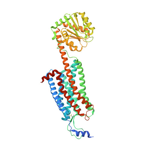
Structure and ligand-binding mechanism of the human OX1 and OX2 orexin receptors.

Yin, J.Babaoglu, K.Brautigam, C.A.Clark, L.Shao, Z.Scheuermann, T.H.Harrell, C.M.Gotter, A.L.Roecker, A.J.Winrow, C.J.Renger, J.J.Coleman, P.J.Rosenbaum, D.M.

(2016) Nat Struct Mol Biol 23: 293-299

  • DOI: https://doi.org/10.1038/nsmb.3183
  • Primary Citation of Related Structures:  
    4ZJ8, 4ZJC

  • PubMed Abstract: 

    The orexin (also known as hypocretin) G protein-coupled receptors (GPCRs) regulate sleep and other behavioral functions in mammals, and are therapeutic targets for sleep and wake disorders. The human receptors hOX1R and hOX2R, which are 64% identical in sequence, have overlapping but distinct physiological functions and potential therapeutic profiles. We determined structures of hOX1R bound to the OX1R-selective antagonist SB-674042 and the dual antagonist suvorexant at 2.8-Å and 2.75-Å resolution, respectively, and used molecular modeling to illuminate mechanisms of antagonist subtype selectivity between hOX1R and hOX2R. The hOX1R structures also reveal a conserved amphipathic α-helix, in the extracellular N-terminal region, that interacts with orexin-A and is essential for high-potency neuropeptide activation at both receptors. The orexin-receptor crystal structures are valuable tools for the design and development of selective orexin-receptor antagonists and agonists.


  • Organizational Affiliation

    Department of Biophysics, University of Texas Southwestern Medical Center, Dallas, Texas, USA.


Macromolecules
Find similar proteins by:  (by identity cutoff)  |  3D Structure
Entity ID: 1
MoleculeChains Sequence LengthOrganismDetailsImage
human OX1R fusion protein to P.abysii glycogen synthase553Homo sapiensPyrococcus abyssi GE5Mutation(s): 1 
Gene Names: HCRTR1PAB2292PYRAB00770
Membrane Entity: Yes 
UniProt & NIH Common Fund Data Resources
Find proteins for Q9V2J8 (Pyrococcus abyssi (strain GE5 / Orsay))
Explore Q9V2J8 
Go to UniProtKB:  Q9V2J8
Find proteins for O43613 (Homo sapiens)
Explore O43613 
Go to UniProtKB:  O43613
PHAROS:  O43613
GTEx:  ENSG00000121764 
Entity Groups  
Sequence Clusters30% Identity50% Identity70% Identity90% Identity95% Identity100% Identity
UniProt GroupsQ9V2J8O43613
Sequence Annotations
Expand
  • Reference Sequence
Small Molecules
Ligands 2 Unique
IDChains Name / Formula / InChI Key2D Diagram3D Interactions
SUV
Query on SUV

Download Ideal Coordinates CCD File 
B [auth A][(7R)-4-(5-chloro-1,3-benzoxazol-2-yl)-7-methyl-1,4-diazepan-1-yl][5-methyl-2-(2H-1,2,3-triazol-2-yl)phenyl]methanone
C23 H23 Cl N6 O2
JYTNQNCOQXFQPK-MRXNPFEDSA-N
OLA
Query on OLA

Download Ideal Coordinates CCD File 
C [auth A]
D [auth A]
E [auth A]
F [auth A]
G [auth A]
OLEIC ACID
C18 H34 O2
ZQPPMHVWECSIRJ-KTKRTIGZSA-N
Binding Affinity Annotations 
IDSourceBinding Affinity
SUV BindingDB:  4ZJ8 Ki: min: 0.4, max: 8.7 (nM) from 6 assay(s)
IC50: min: 50, max: 199 (nM) from 2 assay(s)
Experimental Data & Validation

Experimental Data

  • Method: X-RAY DIFFRACTION
  • Resolution: 2.75 Å
  • R-Value Free:  0.273 (Depositor), 0.280 (DCC) 
  • R-Value Work:  0.223 (Depositor), 0.230 (DCC) 
  • R-Value Observed: 0.225 (Depositor) 
Space Group: P 21 21 21
Unit Cell:
Length ( Å )Angle ( ˚ )
a = 63.242α = 90
b = 64.992β = 90
c = 181.478γ = 90
Software Package:
Software NamePurpose
PHENIXrefinement
Blu-Icedata collection
HKL-3000data scaling
PHASERphasing
HKL-3000data reduction

Structure Validation

View Full Validation Report


Ligand Structure Quality Assessment 

Created with Raphaël 2.3.0Worse 01 BetterLigand structure goodness of fit to experimental dataBest fitted SUVClick on this verticalbar to view detailsBest fitted OLAClick on this verticalbar to view details

Entry History 

Deposition Data

Revision History  (Full details and data files)

  • Version 1.0: 2016-03-09
    Type: Initial release
  • Version 1.1: 2016-03-23
    Changes: Database references
  • Version 1.2: 2016-04-13
    Changes: Database references
  • Version 1.3: 2023-09-27
    Changes: Data collection, Database references, Derived calculations, Refinement description
  • Version 1.4: 2024-10-23
    Changes: Structure summary